Atlas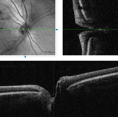
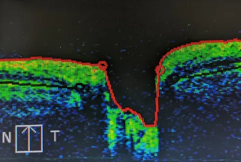
Optic nerve head OCT with scan positions
OCT optic nerveadvancedBaseline disc imaging in a glaucoma suspect; the software shows the en face image and two B scans through the disc.
Diagnosis
Optic nerve head OCT cross sections
- Crosshairs on the en face image mark the vertical and horizontal scan lines
- Nerve fibre layer thickening toward the disc margin
- Cup excavation between the rim edges
- Lamina cribrosa as the bright floor of the cup