Visual atlas
See it before you are asked to name it
132 real images across 7 modality groups. Every one was opened on its Wikimedia Commons file page and checked for what it shows, its licence and whether anyone could be recognised. Tap an image to study it; the practice case behind it grades your localisation and diagnosis.
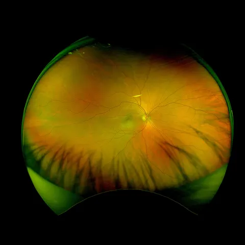 Image of the dayUltra-widefield image at a myopia reviewTry it blind
 132 images
 Colour fundus
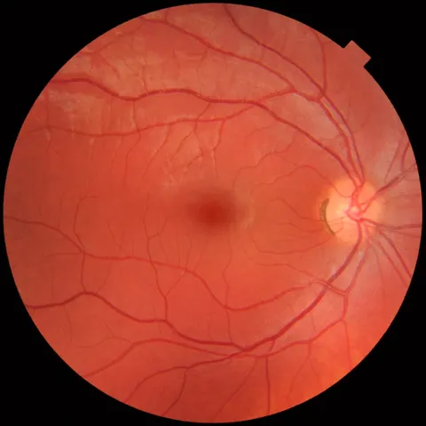
A routine fundus photograph at diabetic screening
A 48-year-old at annual diabetic eye screening, no visual symptoms, acuity 6/6 both eyes.
foundational · comprehensive
 Colour fundus
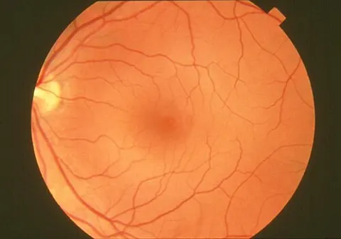
Fundus photograph before a hypertension clinic review
A 55-year-old with treated hypertension, asymptomatic, sent for baseline retinal photography.
foundational · comprehensive
- Colour fundus
Ultra-widefield image at a myopia review
A 29-year-old with high myopia attends for peripheral retinal surveillance after a fellow eye retinal tear.
intermediate · comprehensive
 Colour fundus
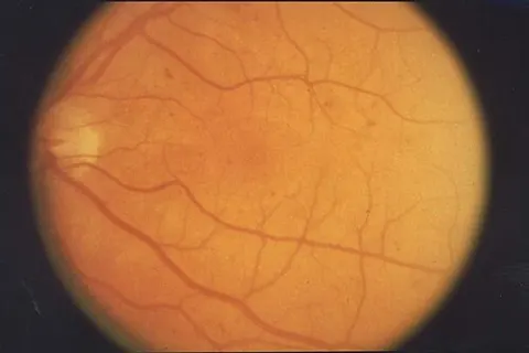
Screening photograph in a patient with type 2 diabetes
A 61-year-old with twelve years of type 2 diabetes and an HbA1c of 74 mmol/mol, no visual symptoms.
foundational · retina
 Colour fundus
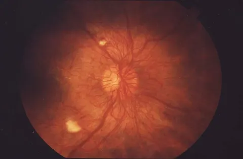
Floaters in a patient with long-standing type 1 diabetes
A 34-year-old with type 1 diabetes since childhood reports new floaters in one eye for a week.
intermediate · retina
 Colour fundusLabelled
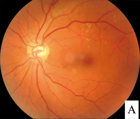
Gradual central blur in a patient with diabetes
A 58-year-old with type 2 diabetes reports three months of gradual blur reading in the left eye.
intermediate · retina
 Colour fundus
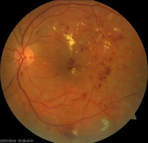
Distortion after previous retinal laser
A 63-year-old with diabetes who had retinal laser two years ago now reports central distortion in the left eye.
intermediate · retina
 Colour fundus
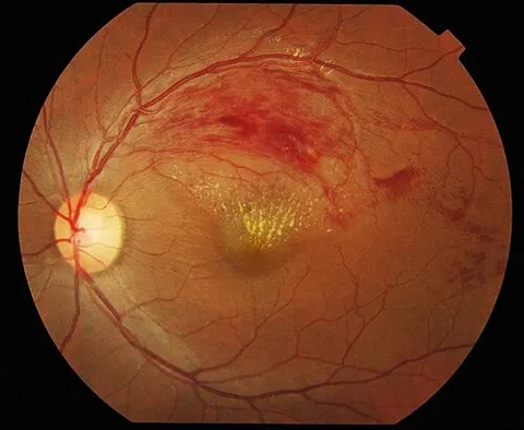
Painless blur in one eye in a hypertensive adult
A 66-year-old with hypertension noticed a blur in the upper part of vision in the left eye over two days.
foundational · retina
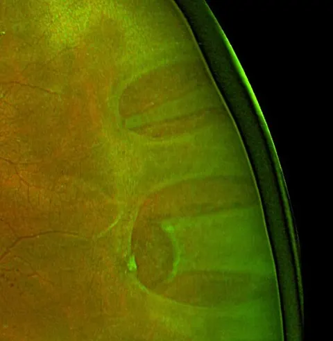 Colour fundus
Flashes, floaters and a shadow in a myope
A 52-year-old myope with a week of flashes and floaters and now a shadow in the nasal field of one eye.
intermediate · retina
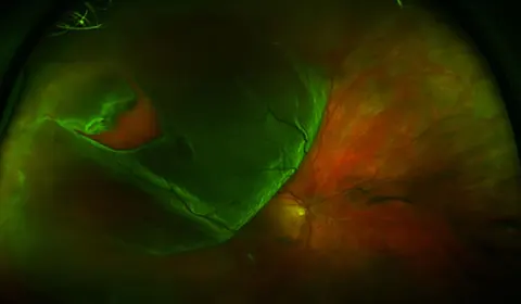 Colour fundus
A curtain over the vision since yesterday
A 47-year-old noticed a dark curtain rising from below in one eye yesterday, preceded by floaters.
foundational · retina
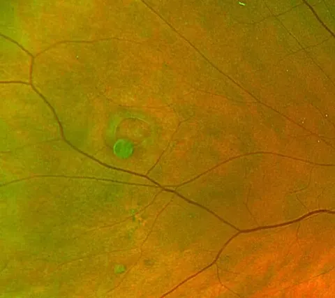 Colour fundus
An incidental finding at a dilated examination
A 38-year-old myope has a dilated examination before refractive surgery; asymptomatic.
intermediate · retina
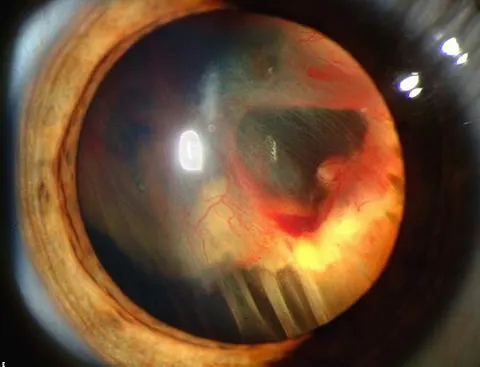 Slit lampLabelled
Progressive visual loss in a young adult with a family history
A 26-year-old with a family history of cerebellar tumours has months of progressive visual loss in one eye.
advanced · retina
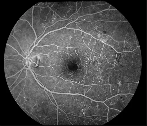 Fluorescein angiography
Angiogram for unexplained central visual loss in diabetes
A 54-year-old with diabetes and acuity of 6/18 whose photograph shows only scattered haemorrhages; the angiogram is requested.
advanced · retina
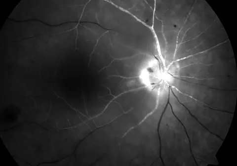 Fluorescein angiography
Sudden painless loss of vision, angiogram frame
A 71-year-old with atrial fibrillation lost vision in one eye two hours ago; this angiogram frame was taken at the arteriovenous phase of the fellow eye.
advanced · retina
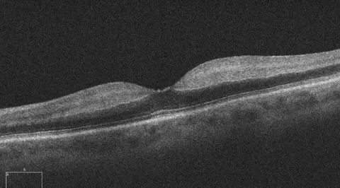 OCT maculaLabelled
Macular OCT hours after sudden visual loss
Same presentation: sudden painless loss of vision in one eye with a relative afferent pupillary defect; OCT of the macula.
advanced · retina
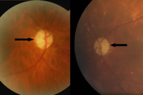 Colour fundusLabelled
Poor colour vision in a young adult with a family history
A 24-year-old with lifelong poor colour vision and slowly declining acuity in both eyes; a parent has the same problem.
intermediate · neuro
 Red-free fundus
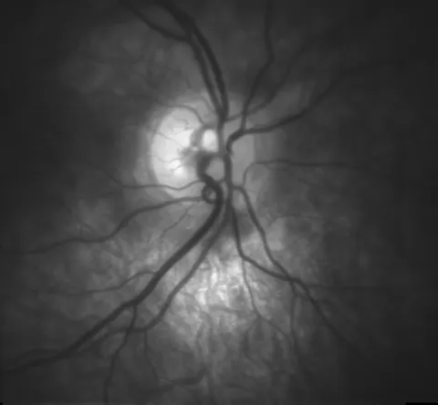
Red-free disc photograph of a glaucoma suspect
A 45-year-old with a family history of glaucoma and pressures of 22 mmHg; red-free disc photography for baseline.
intermediate · glaucoma
 OCT optic nerveLabelled
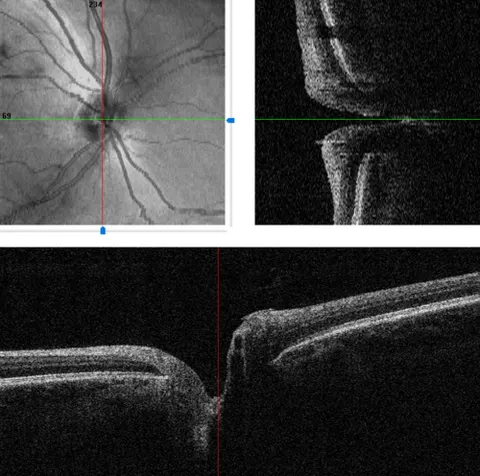
Optic nerve head OCT with scan positions
Baseline disc imaging in a glaucoma suspect; the software shows the en face image and two B scans through the disc.
advanced · glaucoma
 OCT maculaLabelled
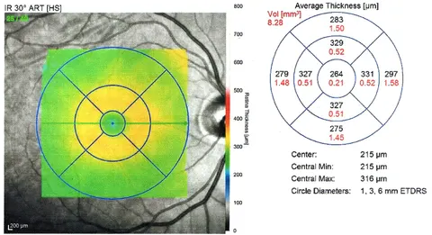
Macular thickness map at baseline
A 50-year-old with diabetes and no retinopathy; baseline macular OCT thickness map of the right eye.
intermediate · retina
 B-scan ultrasound
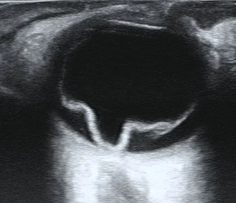
Ultrasound when the fundus cannot be seen
A 60-year-old with sudden loss of vision and a dense vitreous haemorrhage; no fundus view, so a B scan is performed.
intermediate · retina
 B-scan ultrasoundLabelled
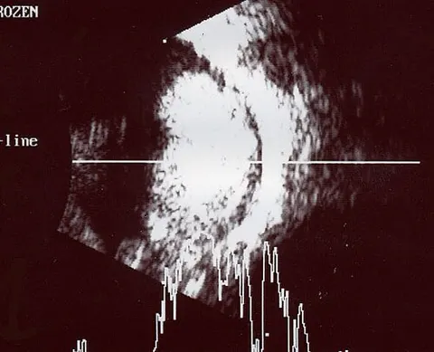
Ultrasound of a child with a white pupil
A two-year-old whose parents noticed a white reflection in one pupil in flash photographs.
advanced · oncology
 External photograph
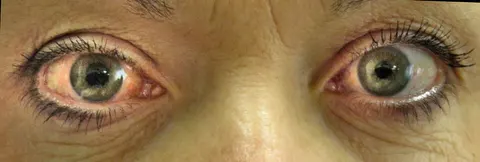
Severe pain, nausea and a red eye in the evening
A 68-year-old long-sighted woman with a night of severe right eye pain, headache, vomiting and haloes around lights.
foundational · glaucoma
 External photograph
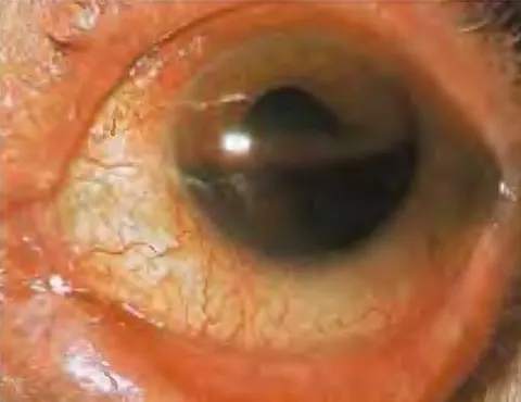
Hit in the eye by a ball
A 19-year-old struck in the eye by a squash ball an hour ago, with pain and blurred vision.
foundational · trauma
 Slit lamp
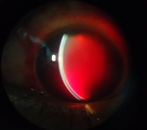
No view of the iris after blunt trauma
A 31-year-old assaulted the previous evening now has profound visual loss in the injured eye.
intermediate · trauma