Guides
Systematic approaches, in depth
Each guide is one method: the pattern to say to yourself at the screen, then every step explained, with the photographs that show it and the labels drawn on the pictures. Read one, then practise it on the cases.
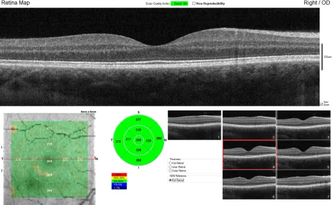
Approach to the OCT macula
A layer-by-layer method for the macular OCT: check the scan, find the fovea, walk from the vitreous to the choroid, name the compartment any fluid sits in, then recognise the pattern.
Read guide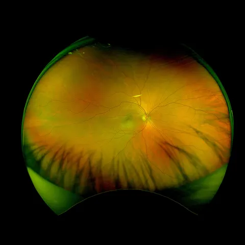
A systematic approach to the fundus
One fixed order for every fundus, at the ophthalmoscope and on the photograph: the view, the disc, the vessels, the macula, then the periphery, with the normal for each and the abnormalities that live there.
Read guide
The red eye
How to sort a red eye in the first two minutes: the three questions that flag the dangerous ones, the pattern of redness that localises the problem, and what to look for at each layer from the lids to the anterior chamber.
Read guide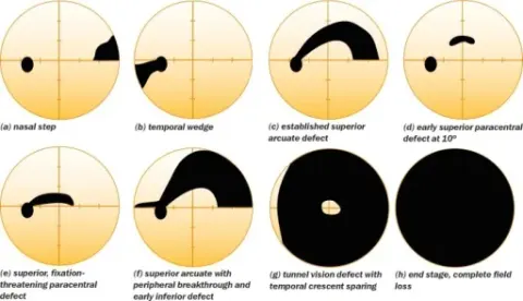
Interpreting visual fields
How to read a visual field: check that the test is reliable, learn what the greyscale and the probability plots show, then use the midlines, the shape of the defect and its match across the two eyes to place the lesion from the retina to the occipital cortex.
Read guide