Atlas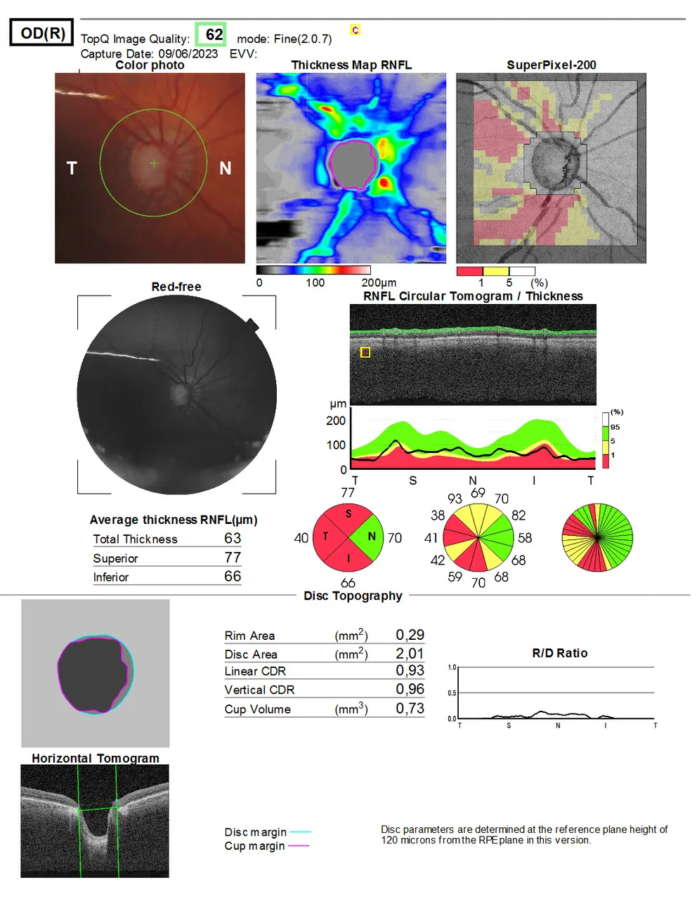
The optic nerve head report of a right eye with glaucoma
OCT optic nerveadvancedThe right eye of an 80-year-old man under treatment for glaucoma, imaged at review.
A full instrument report; the panels are labelled at source.
Diagnosis
Glaucomatous optic neuropathy on optic nerve head imaging
- Retinal nerve fibre layer thickness map thinned in the inferior and superior sectors
- Sector chart flagged red and yellow where the thickness is outside normal limits
- Large cup with a thin rim on the disc topography
- Cup to disc ratio recorded above 0.9