Atlas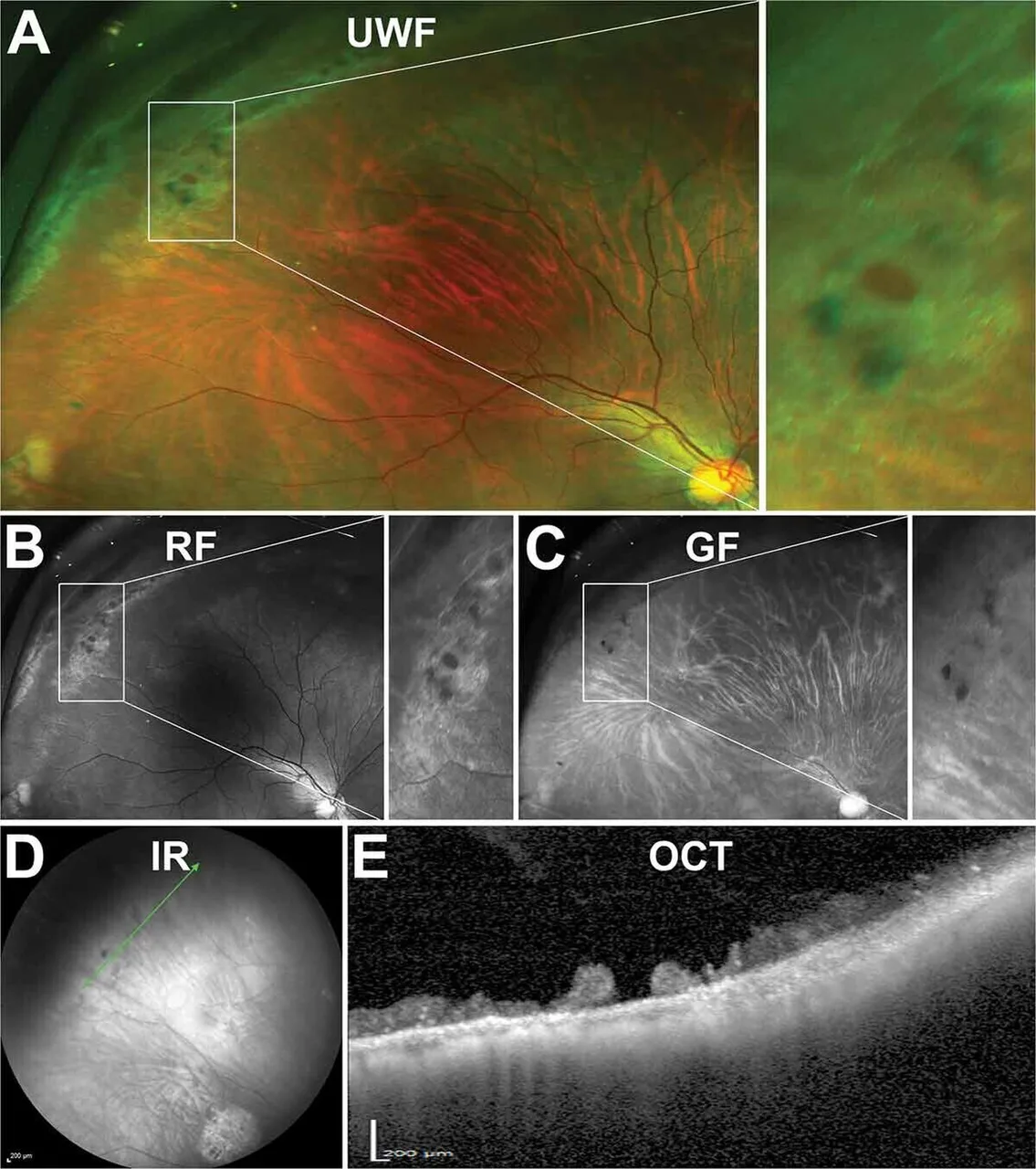
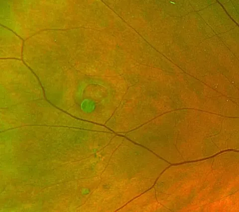
An atrophic hole within lattice, on every modality
Colour fundusadvancedA 23-year-old myopic woman with no symptoms, found at a routine dilated examination to have a peripheral lesion.
Multi-panel figure with the source's panel labels.
Diagnosis
Atrophic retinal hole within lattice degeneration
- Ultra-widefield image with a peripheral patch of lattice degeneration
- Red-free and green-filter images showing the criss-cross lines of lattice
- Infrared image of the area
- OCT through the lesion showing the full thickness hole with overlying vitreous condensation