Atlas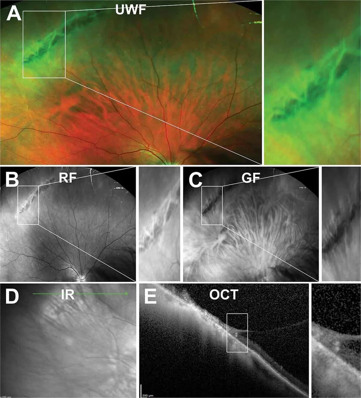
Criss-cross white lines in the far periphery of a 60-year-old
Colour fundusadvancedA 60-year-old East Asian myope with no symptoms, found at a dilated examination to have a peripheral lesion in the superotemporal retina.
Multi-panel figure with the source's panel labels.
Diagnosis
Lattice degeneration
- Sharply bordered, spindle-shaped area of thinned retina parallel to the ora
- Criss-cross white lines of sclerosed vessels within it
- Pigment clumps
- OCT showing the thinned retina with firm vitreous attachment at its edges