Extraocular muscles · 14 of 24
What does the superior rectus do?
The superior rectus runs forward from the annulus of Zinn at the orbital apex, over the top of the globe, to insert on the sclera about eight millimetres behind the upper limbus. Because the orbit points outwards and the muscle runs straight back, it pulls at an angle to the eye's axis, so it elevates most purely when the eye is turned out, and it also intorts and adducts.
- Action
- Elevation; also intorsion and adduction
- Purest test
- Looking up with the eye abducted
- Nerve
- Superior division of the oculomotor nerve (CN III)
- Paired with
- The inferior oblique of the other eye, in upgaze
Why the actions are what they are
The muscle's line of pull runs from the apex, which lies medial to the eye, to the top of the globe in front, so it makes an angle of about twenty three degrees with the visual axis when the eye looks straight ahead. Its force therefore has three components: up, in, and a rotation of the top of the eye towards the nose. Turn the eye out by twenty three degrees and the muscle lines up with the axis: now it only elevates. That is why the examiner tests each muscle in the position where it is the main mover.
It shares a fascial sheath with the levator of the upper lid above it, which is why the lid rises when the eye looks up, and why surgery on the superior rectus can change the lid height.
When it fails
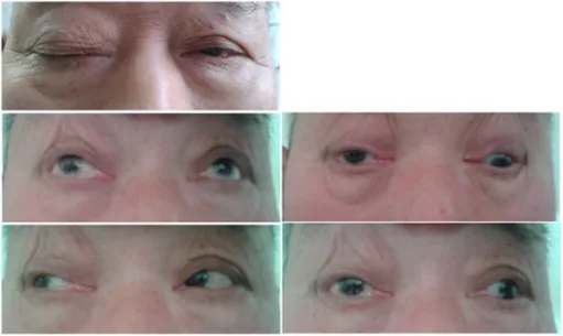
A third nerve palsy takes the superior rectus with the medial and inferior recti, the inferior oblique and the levator: the eye sits down and out under a drooping lid. Thyroid eye disease thickens and stiffens the inferior rectus first and the superior rectus after it, restricting the eye rather than weakening it, so a forced duction test finds the eye stuck. In myasthenia any muscle can weaken and the pattern changes through the day.
Examine it
Tested with the eye turned out and up.
Ask OphthoSpace about the superior rectus
AI responses are for education and may contain errors. OphthoSpace is not for primary clinical diagnosis.