Learn / Neuro-ophthalmology
Neuro-ophthalmology
Localise the loss. Read the pupil. Name the nerve.
The eye is an outpost of the brain, and the optic nerve, the pupils and the eye movements are the three ways the brain shows itself at the slit lamp. Neuro-ophthalmology is localisation: where along the pathway, and how urgently.
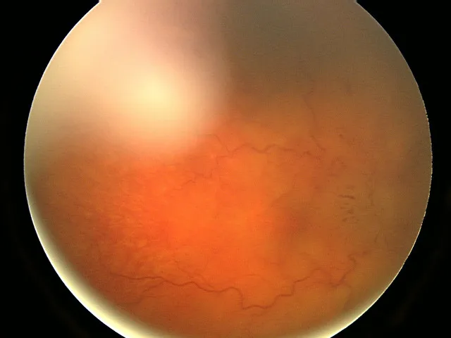
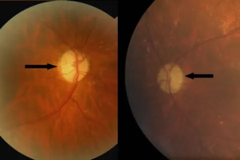
Headache worse on waking, and a photograph of the right discOpen in the atlas
The optic nerve and its diseases
One million ganglion cell axons, myelinated behind the lamina cribrosa, wrapped in meninges. Damage anywhere along it shows as reduced acuity, reduced colour vision, a relative afferent pupillary defect and a field defect; the disc may be swollen, normal or pale depending on where and when.
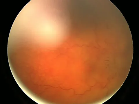
- A verified photograph of papilloedema is not yet in the atlas; the pale disc of atrophy is.
| Condition | Who and how | Disc | The key point |
|---|---|---|---|
| Optic neuritis | Young adult, days of blur, pain on eye movement, RAPD, colour loss | Normal (retrobulbar) or swollen | Often the first event of multiple sclerosis; MRI of the brain; recovers over weeks |
| Anterior ischaemic optic neuropathy | Over 50, sudden painless loss on waking, altitudinal field | Swollen, then pale | Non-arteritic in a crowded disc; ARTERITIC in giant cell arteritis: check ESR and CRP the same day |
| Papilloedema | Both discs swollen from raised intracranial pressure; headache worse lying, transient obscurations, a sixth nerve palsy | Swollen, both sides, vision preserved early | Imaging the same day; then lumbar puncture if imaging is clear |
| Compressive neuropathy | Slow loss, sometimes proptosis | Normal or pale | Image the orbit and chiasm |
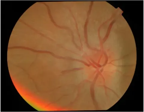
| Optic atrophy | The end state of any of the above | Pale, sharp margins | Find the cause: the history says when it happened |
Giant cell arteritis
Emergent · nowA vasculitis of the over-fifties: temporal headache, scalp tenderness, jaw claudication, malaise, weight loss and polymyalgia, with a raised ESR and CRP and a thickened, tender, pulseless temporal artery. It causes arteritic ischaemic optic neuropathy, sudden and severe, and takes the second eye within days if untreated. Steroids start on suspicion, before the temporal artery biopsy, because the biopsy stays positive for weeks and the eye does not wait.
The visual pathway and the field
Retina, nerve, chiasm, tract, radiations, cortex: the field defect tells you which. One eye means in front of the chiasm; respecting the vertical midline means the chiasm or behind; homonymous means behind, on the opposite side; congruity increases posteriorly; macular sparing is occipital. The Visual Field Lab drills every pattern.
The pupils
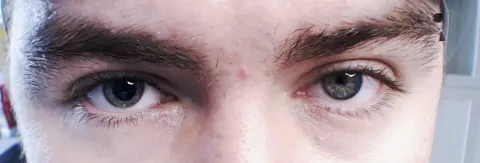
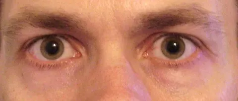
The afferent limb runs with the optic nerve to the pretectum and both Edinger-Westphal nuclei; the efferent parasympathetic limb runs with the third nerve; the sympathetic runs a three-neuron path through the neck. The swinging light finds the afferent defect; anisocoria in the light versus the dark separates a third nerve or tonic pupil from a Horner syndrome; and a dilated pupil with a painful third nerve palsy is an aneurysm until proven otherwise.
Cranial nerve palsies and diplopia
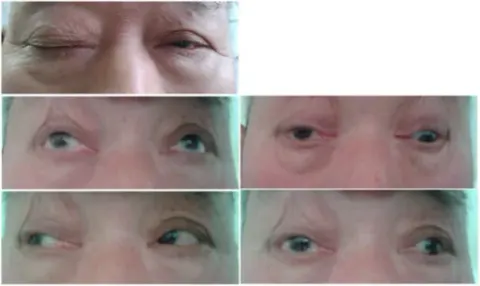
Binocular diplopia that goes when either eye is closed is a misalignment: a third, fourth or sixth nerve palsy, a muscle problem (thyroid eye disease, myasthenia, an orbital floor fracture) or an internuclear ophthalmoplegia. The direction of the double vision and the position of gaze in which it is worst name the muscle, and the muscle names the nerve. Monocular diplopia that persists with one eye is optical: astigmatism, cataract, a dry eye.
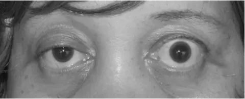
Horner syndrome
Ptosis, miosis and anhidrosis on one side, from a break in the sympathetic chain: the brainstem and cervical cord (stroke, demyelination, syringomyelia), the lung apex and neck (a Pancoast tumour, thyroid surgery), or the carotid (dissection, which is painful and needs the vessel imaged the same day). The pupil difference is greater in the dark and the affected pupil dilates slowly.
Go and look
Where this topic leads