Examine / Colour vision
Colour vision
The optic nerve's early warning, and the inherited difference that is not disease.
Colour vision is tested to find inherited red-green deficiency, common and lifelong, and to find acquired loss, which is an early sign of optic nerve disease and is compared between the two eyes.
Inherited red-green deficiency
About one in twelve men and one in two hundred women, X-linked: a missing or shifted long-wavelength (protan) or middle-wavelength (deutan) cone pigment. Both eyes, present from birth, stable, and not a disease; it matters for a few occupations and for reading colour-coded charts. Blue-yellow (tritan) deficiency is rare and is more often acquired.
Acquired colour loss
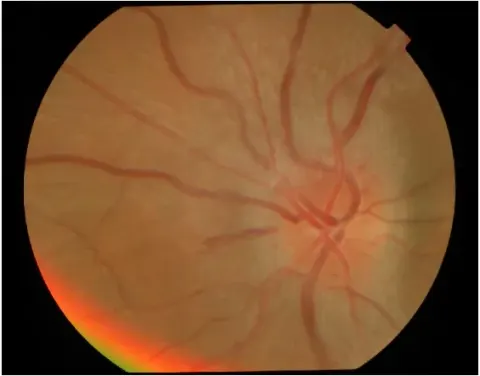
Optic nerve disease takes colour early and asymmetrically: the eye with optic neuritis sees red as washed out (red desaturation), and this is tested at the bedside by showing a red target to each eye in turn and asking which is redder. Ganglion cell and nerve disease tend to take red-green; retinal disease and cataract tend to take blue-yellow (Köllner's rule, a tendency, not a law). Drugs matter: ethambutol and, rarely, hydroxychloroquine.
The tests
Ishihara plates screen for red-green deficiency: numbers or paths in dots of confusable colours, read at reading distance in good light, each eye in turn, with the score recorded as plates correct out of the number shown. They do not test blue-yellow. The Farnsworth D-15 and the 100-hue arrange coloured caps in order and classify the type and severity. The Ishihara plates are a published, copyrighted test and are not reproduced here; the principle is what a student needs, and the plates are in every eye clinic.
- The first Ishihara plate is a control that anyone with sight can read; a patient who fails it is not trying, or cannot see the plate at all.
Go and look
Where this topic leads