Examine / External eye
The external eye
Look before you magnify.
Most of the external examination is done from across the room and with a torch: the lids, the lashes, the position of the eyes, the conjunctiva, and the one manoeuvre students avoid and should not, everting the upper lid.
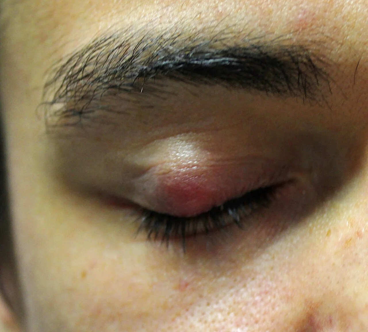
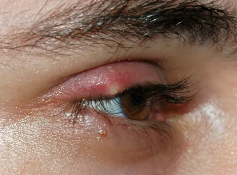
A lump in the eyelid for three weeksOpen in the atlas
From across the room
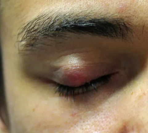
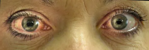
Symmetry. Are the eyes level and aligned? Is one lid lower (ptosis), or higher (retraction)? Is one eye forward (proptosis: look from above and behind)? Is there swelling, redness, a lump, a rash in a dermatome? Is the head tilted or turned? A surprising amount of neuro-ophthalmology is visible from the doorway.
The lids and lashes
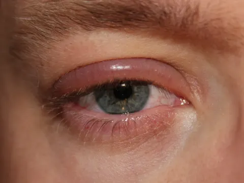
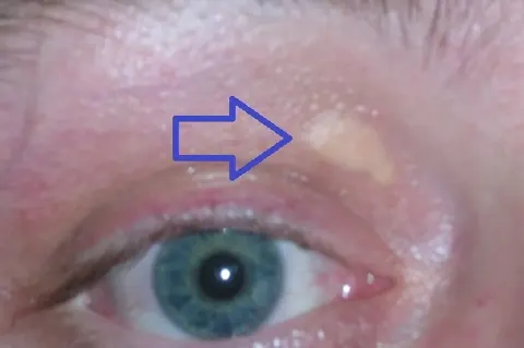
The margin: crusting and collarettes at the lash bases (anterior blepharitis), capped meibomian orifices and a foamy tear film (posterior blepharitis), lashes turning in (trichiasis) or a lid turning in (entropion) or out (ectropion). Lumps: a chalazion in the tarsus, a stye at a follicle, and the pearly, telangiectatic, ulcerated lump with lost lashes that is a basal cell carcinoma. Measure a ptosis: the distance from the corneal light reflex to the upper lid margin (normally 4 to 5 mm) and the levator function (lid excursion from downgaze to upgaze with the brow held).
Everting the upper lid
Ask the patient to look down. Hold the lashes of the upper lid between finger and thumb, pull the lid gently down and away from the eye, place a cotton bud on the lid a centimetre above the margin, and fold the lid up over it. The tarsal conjunctiva is now in view: the subtarsal foreign body that scratches the cornea with every blink, the giant papillae of a contact lens wearer, the follicles of chlamydia. Let go and the lid returns on its own with an upward look.
- Every red, painful eye with a foreign body sensation has its upper lid everted. A cornea with vertical linear scratches has a foreign body under the lid until the lid is looked at.
The conjunctiva and sclera
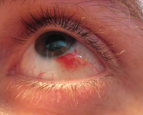
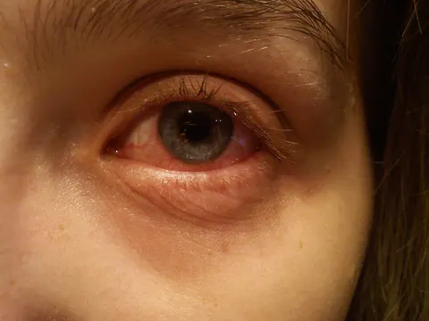
Pull the lower lid down and look at the fornix and the bulbar surface; ask the patient to look up, down and to each side. Redness: its pattern (diffuse, ciliary, sectoral), its depth (superficial vessels move with the conjunctiva and blanch; deep ones do not), and what is in the fornix (pus, follicles, papillae, a foreign body). A flat bright red patch is a subconjunctival haemorrhage; a fleshy wedge onto the nasal cornea is a pterygium.
Go and look
Where this topic leads