Learn / Oculoplastics and orbit
Oculoplastics and the orbit
Lids, lashes, tears, and the space behind the eye.
The adnexa protect the eye and keep it wet, and the orbit holds it. Most of what goes wrong here is seen from across the room: a lump on the lid, a lid that droops or turns, an eye that bulges.
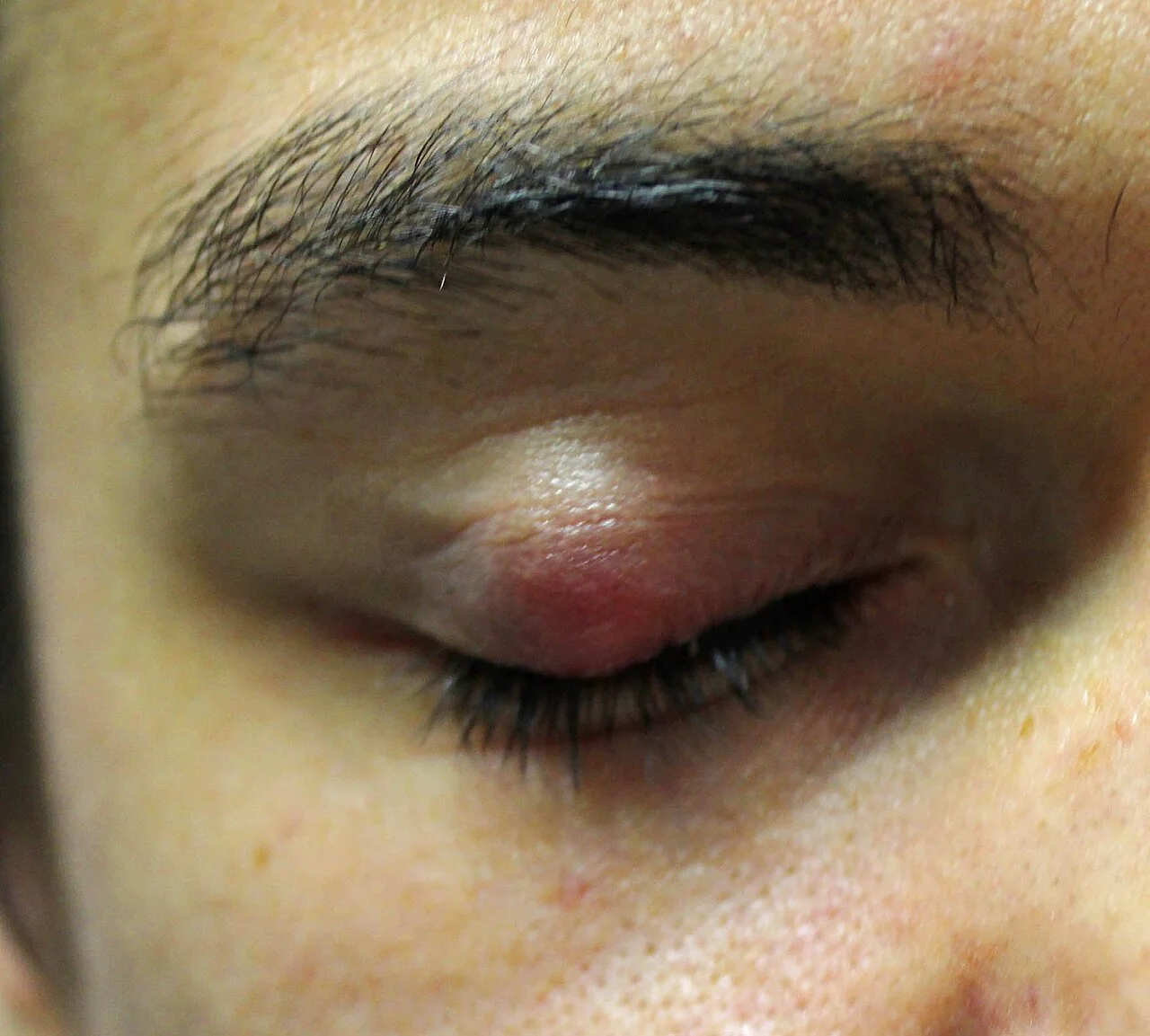
A lump in the eyelid for three weeksOpen in the atlas
Lid lumps
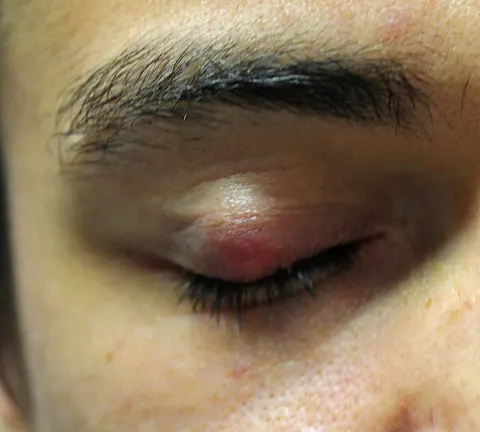
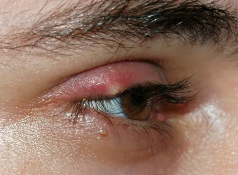
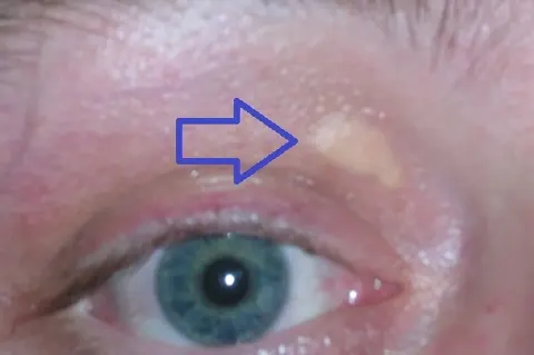
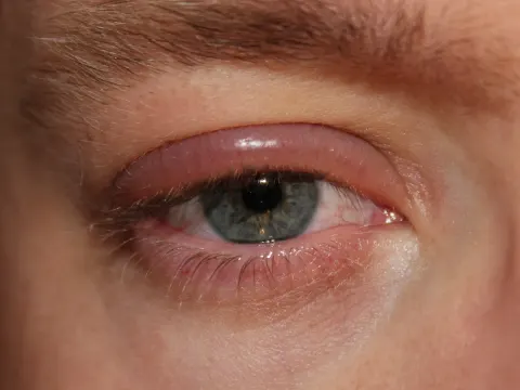
A chalazion is a blocked meibomian gland in the tarsal plate: a firm, painless lump that points to the inside of the lid, after an acute phase that can be red and tender. A hordeolum (stye) is an infected lash follicle at the lid margin, tender and pointing outwards. Both settle with warm compresses; a chalazion that persists is incised from the inside. A lid lump that is pearly, ulcerated, loses its lashes or recurs is a basal cell carcinoma until the histology says otherwise.
Lid position
Ptosis is a drooping upper lid: aponeurotic with age (the commonest), congenital, mechanical, or neurogenic, where it matters most: a third nerve palsy (complete ptosis, the eye down and out, a big pupil) or a Horner syndrome (mild ptosis, a small pupil). Myasthenia gives a fatigable ptosis that varies through the day. Entropion turns the lid in so the lashes scratch the cornea; ectropion turns it out so the eye waters and the exposed conjunctiva reddens. Lid retraction, showing sclera above the cornea, is thyroid eye disease until proven otherwise.
Blepharitis and dry eye
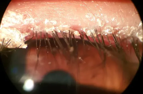
Chronic inflammation of the lid margins, anterior (staphylococcal crusting and collarettes at the lash bases) or posterior (meibomian gland dysfunction, capped orifices, a foamy tear film). Gritty, burning, red-rimmed eyes, worse in the morning, with recurrent chalazia. Lid hygiene, warm compresses and lubricants are the treatment, for life. Dry eye follows: an unstable tear film, punctate staining of the lower cornea, and symptoms worse with screens and in the wind.
Tears and their drainage
The lacrimal gland in the superotemporal orbit makes the aqueous layer; the meibomian glands make the lipid layer that stops it evaporating; the goblet cells make the mucin that spreads it. Tears drain through the puncta, the canaliculi and the sac into the nasolacrimal duct under the inferior turbinate. A watering eye is too much (irritation) or too little drainage (a blocked duct, an out-turned punctum); dacryocystitis is an infected sac, a red tender swelling below the medial canthus.

The orbit: proptosis and cellulitis
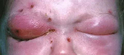
Urgent · same dayProptosis is the eye pushed forward: thyroid eye disease (the commonest cause in adults, with lid retraction, lid lag and restricted upgaze), an orbital tumour, a vascular lesion, or inflammation. Measured with an exophthalmometer, imaged with CT. Orbital cellulitis, from adjacent sinusitis, is proptosis with a red, swollen lid, painful restricted eye movements, fever and, when it threatens the nerve, reduced vision and an afferent pupillary defect: admission, intravenous antibiotics and CT the same day. Preseptal cellulitis stops at the septum: lid swelling alone, with normal movement and vision.
Go and look
Where this topic leads