Examine / Fluorescein staining
Fluorescein staining
The dye that shows where the epithelium is missing, and whether the eye is leaking.
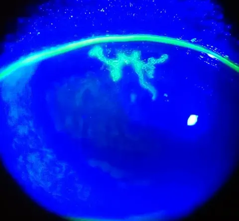
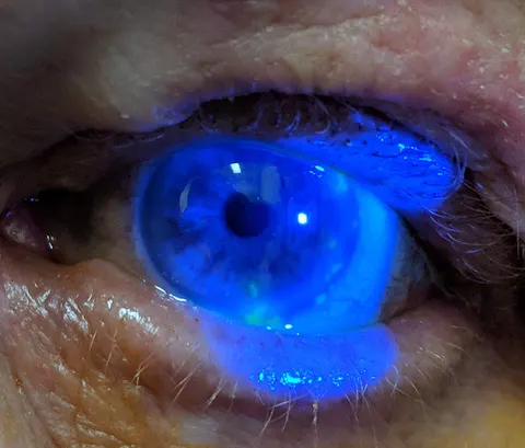
Fluorescein is an orange dye that glows green under cobalt blue light and pools wherever the corneal or conjunctival epithelium is absent. A drop and a blue light turn an invisible abrasion into a bright map, show the branching of a herpetic ulcer, time the tear film, and reveal a leaking wound with the Seidel test.
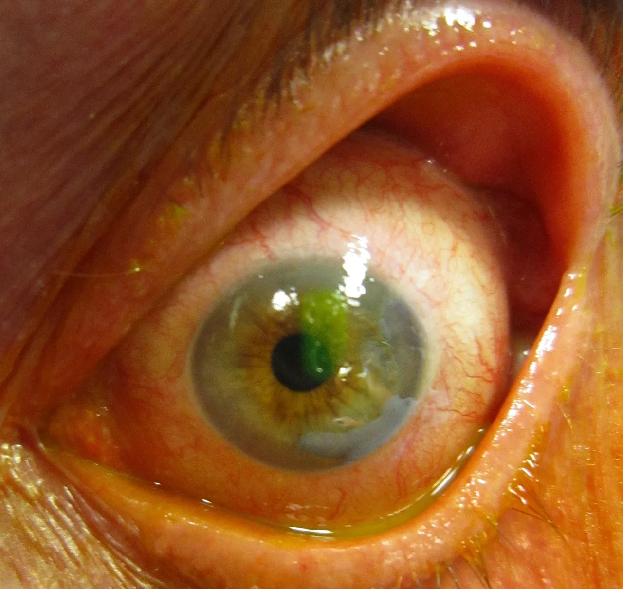
Scratched by a fingernail this morning, under the blue lightOpen in the atlas
How it is done
Take out contact lenses first: soft lenses take up the dye permanently. Wet a fluorescein strip with a drop of saline or topical anaesthetic, touch it to the inside of the lower lid without touching the cornea, and ask the patient to blink a few times to spread it. Then switch the slit lamp to the cobalt blue filter, open the beam wide and survey the whole cornea and conjunctiva at low magnification before going in closer; a yellow barrier filter in front of the eyepiece makes the green stand out from the blue.
Without a slit lamp, the blue light on a direct ophthalmoscope or a pen torch with a blue filter does the same job at the bedside, and the finding is the same: intact epithelium does not stain, and anything that glows bright green is a place where it has gone.
- Too much dye floods the eye and hides small defects; a small amount, then blink, then look.
- Warn the patient that their tears and the inside of their nose will be yellow for a few hours.
- Evert the upper lid whenever there are fine vertical scratches on the cornea: the foreign body is usually under it.
What the patterns mean
The shape and place of the staining is the diagnosis more often than not.
| Pattern | Means | Do |
|---|---|---|
| A sharp-edged patch of bright green with clear cornea around it | Corneal abrasion | Look for the cause, check under the lid, antibiotic ointment, no patch or steroid, review if not healed in a day or two |
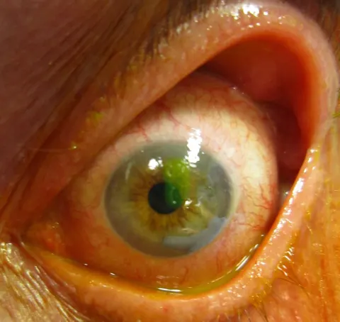
| A branching line with knobbed ends | Dendritic ulcer of herpes simplex | Antiviral, no steroid without an ophthalmologist; the steroid turns a dendrite into a geographic ulcer |
| A raised, stuck-on branching lesion without terminal bulbs | Pseudodendrite of herpes zoster | Treat the zoster; the rash on the nose tip means the eye is involved |
| Fine dots scattered across the lower cornea or between the lids | Punctate epithelial erosions: dry eye, exposure, drop toxicity, contact lens overwear | The distribution tells the cause; lubricants, and stop the offending drop or lens |
| Fine vertical scratches in the upper cornea | A foreign body under the upper lid | Evert the lid and remove it |
| A white infiltrate that takes up dye, in a contact lens wearer | Infectious keratitis until proven otherwise | Same-day ophthalmology, scrape before antibiotics, no steroid |
The Seidel test
Emergent · nowWhen a full-thickness wound is possible, after trauma or surgery, paint concentrated fluorescein from the strip directly over the suspect site and watch under cobalt blue. Aqueous leaking through the wound dilutes the dye into a bright green stream that runs down the cornea: a positive Seidel test, which means the globe is open. Stop examining, put a shield over the eye, give no more drops and no pressure, and refer as an emergency.
Tear break-up time
With a small amount of fluorescein in the tear film, ask the patient to blink and then hold the eye open, and count the seconds until the first dark dry spot appears in the green film. Under about ten seconds is an unstable tear film, the objective sign of evaporative dry eye. It is quick, and it pairs with the punctate staining pattern to explain a gritty, burning eye.
Go and look
Where this topic leads