Learn / Anterior segment
Anterior segment
Cornea, conjunctiva, iris, chamber, lens.
Everything the slit lamp sees first. The anterior segment is where most red eyes live, where a contact lens does its damage, and where the pressure is made and drained.
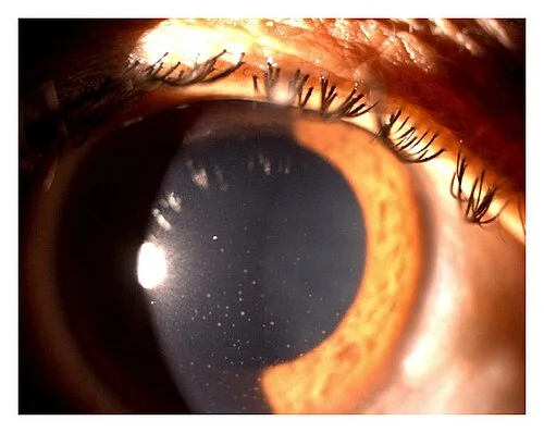
Photophobia and a small pupilOpen in the atlas
The cornea
Half a millimetre of transparent, avascular tissue, fed by the tears in front and the aqueous behind, and the most densely innervated surface in the body, which is why a scratch on it hurts the way it does. Five layers: epithelium (which heals in a day or two without a scar), Bowman's layer, the stroma (nine tenths of the thickness; it scars), Descemet's membrane, and the endothelium (a single layer that pumps the stroma dry and does not regenerate).
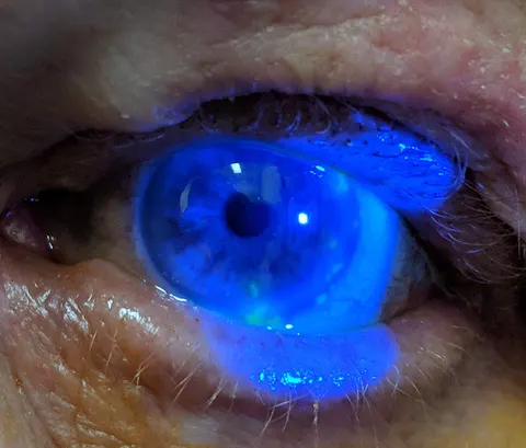
Epithelial defects take up fluorescein and glow green under cobalt blue light: an abrasion, a dendrite, punctate keratopathy. A stromal infiltrate is a white opacity, and with an overlying epithelial defect it is an ulcer. A hazy, thick cornea is oedema, from a failing endothelium or a pressure spike.
- Abrasion: pain, photophobia, a fluorescein-staining defect, a clear stroma; heals in 24 to 72 hours.
- Bacterial keratitis: a contact lens wearer, a white infiltrate with an epithelial defect, sometimes a hypopyon; same-day scrape and intensive antibiotics.
- Herpes simplex keratitis: a branching dendrite with terminal bulbs on fluorescein; reduced corneal sensation; topical antiviral, and never steroid alone.
- Herpes zoster ophthalmicus: the ophthalmic dermatome, the tip of the nose (Hutchinson's sign) marking nasociliary involvement, pseudodendrites, uveitis, and complications for months.
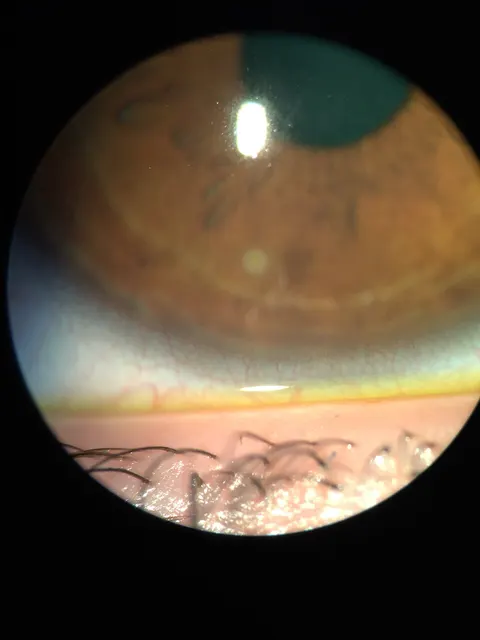
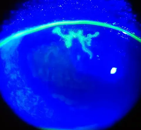
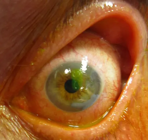
The conjunctiva
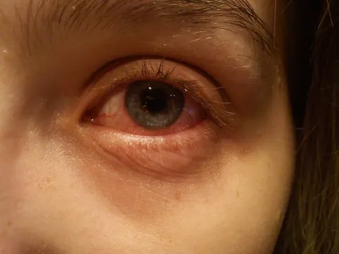
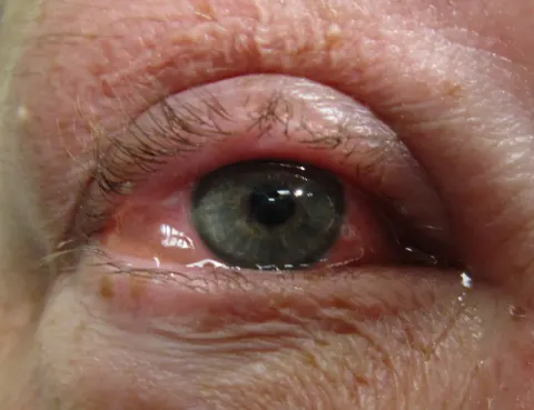
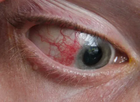
The thin vascular membrane over the sclera (bulbar) and the inside of the lids (tarsal), meeting in the fornices. Its injection pattern is the first thing to read in a red eye: diffuse, deepest in the fornices, in conjunctivitis; around the limbus (ciliary flush) in keratitis and uveitis; sectoral in episcleritis.
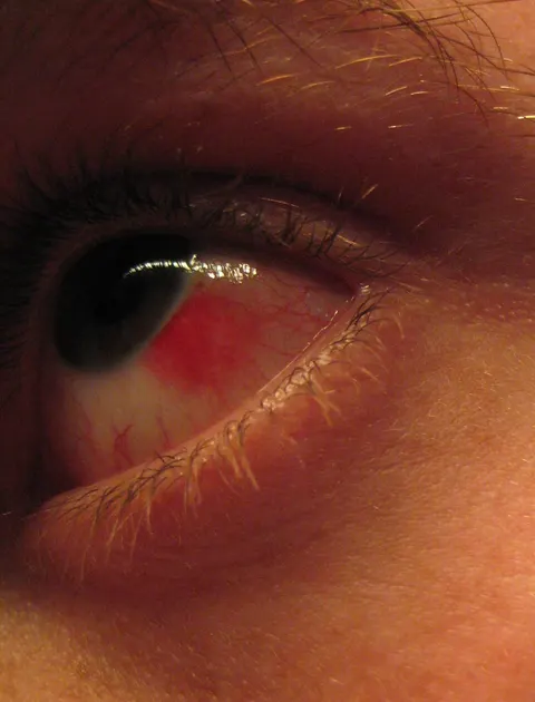
Follicles (pale, round, lymphoid bumps in the fornix) mean a viral or chlamydial cause; papillae (red, vascular cores, on the tarsal surface) mean allergy or a chronic irritant. A flat, bright red, sharply bordered patch is a subconjunctival haemorrhage, alarming and harmless.
- Viral conjunctivitis: watery discharge, follicles, a tender preauricular node, often after a cold; contagious; self-limiting.
- Bacterial conjunctivitis: purulent discharge, lids stuck in the morning; topical antibiotic shortens it.
- Allergic: itch above all, chemosis, papillae, both eyes.
- Pterygium: a wing of conjunctiva growing onto the nasal cornea in sun-exposed eyes.
The anterior chamber
The aqueous-filled space between cornea and iris. Aqueous is secreted by the ciliary processes into the posterior chamber, flows through the pupil, and drains at the angle through the trabecular meshwork into Schlemm's canal. Judge its depth at the slit lamp (a shallow chamber is a risk for angle closure), then look into it with a short, bright, narrow beam in a dark room.
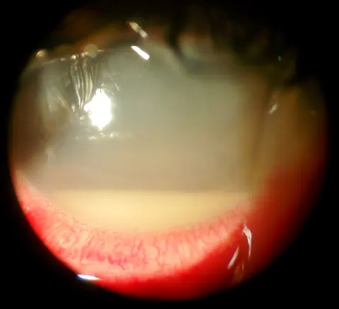
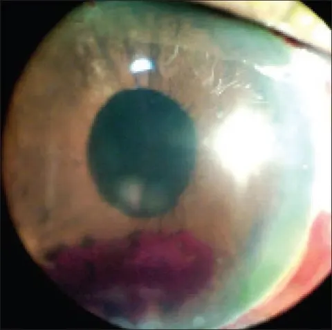
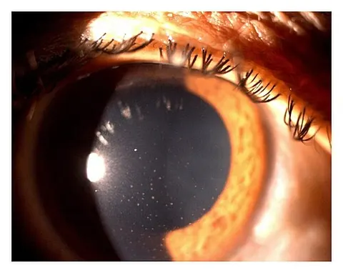
Cells are white blood cells drifting in the beam like dust in sunlight; flare is the beam becoming visible in the aqueous because protein has leaked into it. Both are graded, and both mean inflammation: anterior uveitis. What settles below tells the diagnosis: white cells layer as a hypopyon, blood as a hyphaema.
- Hypopyon: a level of pus in the chamber; with a corneal ulcer it is infectious keratitis, without one it may be uveitis, and after surgery it is endophthalmitis until proven otherwise.
- Hyphaema: blood after blunt trauma; a marker of a hard blow, with the risks of rebleed, a pressure rise and an open globe behind it.
- A shallow chamber with a hard eye and a fixed mid-dilated pupil is acute angle closure.
The iris and anterior uveitis
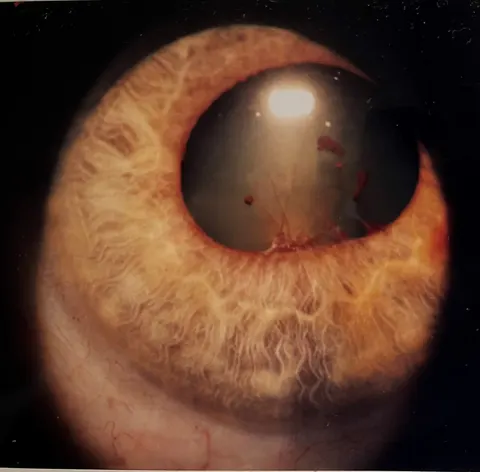
The iris, ciliary body and choroid together are the uvea, and inflammation of the front of it is anterior uveitis (iritis). The patient has a deep ache, photophobia and a red eye with ciliary flush, and at the slit lamp: cells and flare, keratic precipitates on the corneal endothelium, a small pupil from sphincter spasm, and, if it has gone on, posterior synechiae sticking the iris to the lens so that the pupil dilates unevenly.
Half of cases are HLA-B27 associated (ankylosing spondylitis, reactive arthritis, inflammatory bowel disease, psoriasis); some are sarcoidosis, herpetic, or idiopathic. Treatment is topical steroid to quell the inflammation and a cycloplegic to relieve the spasm and break the synechiae, with the pressure checked because both the disease and the steroid can raise it.
The lens and cataract
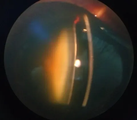
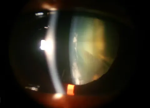
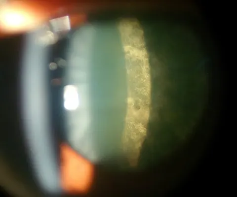
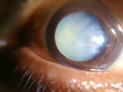
The lens grows throughout life, laying down fibres it never sheds, and with age it yellows, stiffens (presbyopia in the forties) and clouds. Cataract is any opacity in it: nuclear sclerosis (a myopic shift and dulled colours), cortical spokes (glare), posterior subcapsular plaque (glare and near vision, in steroid users, diabetics and the young). Gradual painless loss of vision with a dimmed red reflex is the picture.
Cataract surgery (phacoemulsification with an intraocular lens) is the most common operation performed. Its rare emergency is endophthalmitis in the first week: pain, a hypopyon and falling vision after surgery need the same day.
- No verified photograph of a cataract at the slit lamp is in the atlas yet; the lens is taught here in words and on the 3D eye until one is found.
Go and look
Where this topic leads
- Slit Lamp LabExamine the anterior segment on real photographs
- The red eye pathwaySort the causes by the questions that separate them
- Atlas: slit lamp and externalEvery anterior segment image
- The rash that came first: zoster in DermSpaceOpen in DermSpace
- Topical steroids, antibiotics and cycloplegicsOpen in PharmSpace