Learn / Optics and the lens
Optics, refraction and the lens
Why the image lands where it lands.
The cornea does two thirds of the focusing and the lens does the rest, and adjusts. When the image misses the retina the vision blurs; the pinhole tells you whether blur is optical, and the lens's clouding is cataract.
The eye as an optical system
About 60 dioptres of power: roughly 43 from the cornea (air to tissue is the big change of refractive index) and 17 to 20 from the lens, in an eye 24 mm long. An emmetropic eye focuses distant light on the retina with the lens relaxed. The refractive errors are mismatches between power and length.
| Error | Where the image lands | The eye | Corrected with |
|---|---|---|---|
| Myopia (short sight) | In front of the retina | Too long, or too powerful | A minus (concave) lens |
| Hyperopia (long sight) | Behind the retina | Too short, or too weak; the young eye accommodates to hide it | A plus (convex) lens |
| Astigmatism | Two focal lines, not one point | A cornea curved more in one meridian than the other | A cylinder at an axis |
| Presbyopia | Near objects behind the retina | A stiff lens that can no longer accommodate, from the mid-forties | Reading glasses (plus for near) |
Accommodation
To focus near, the ciliary muscle contracts, the ring of muscle narrows, the zonules slacken and the elastic lens rounds up, adding power. Parasympathetic, through the third nerve, along with the pupil's constriction and the eyes' convergence: the near triad. The lens stiffens with age and the range of accommodation shrinks from about 14 dioptres in childhood to nearly nothing by sixty: presbyopia.
The lens and zonules on the 3D eye The ciliary body on the 3D eye
Reading a prescription
Sphere (the spherical power, minus for myopia and plus for hyperopia), cylinder (the astigmatic correction) and axis (the meridian of the cylinder, 0 to 180 degrees), for each eye, with an add for near in presbyopia. A prescription of minus 3.00 is a moderate myope; plus 2.00 add 2.50 is a presbyopic hyperope.
The pinhole
A pinhole admits only the central rays, which need little focusing, so vision that improves through a pinhole is blurred by refractive error and will improve with glasses. Vision that does not improve is limited by something else: the media (cataract), the retina or the nerve. It is the single most useful thing to do with a reduced acuity.
Cataract
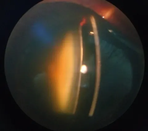
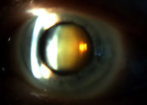
Any opacity of the lens. Nuclear sclerosis yellows and hardens the centre and shifts the eye towards myopia (the elderly patient who no longer needs reading glasses); cortical cataract makes radial spokes and glare; posterior subcapsular cataract sits at the back of the lens in the visual axis and ruins reading and night driving early, in diabetics, steroid users and after uveitis. Painless, gradual, with a dulled red reflex. Phacoemulsification with an intraocular lens restores vision in a day; the complications to know are posterior capsule opacification (lasered), and endophthalmitis (an emergency).
Go and look
Where this topic leads