Adnexa · 24 of 24
What is the lower tarsal plate?
The lower tarsal plate is the smaller of the two, about four millimetres tall, holding the shape of the lower lid and its row of meibomian glands. The lower lid does not lift; it is held against the eye by the canthal tendons and the orbicularis, and it is its position rather than its movement that fails with age.
- Height
- About 4 mm
- Contains
- About 20 to 25 meibomian glands
- Held by
- The medial and lateral canthal tendons, and the tone of the orbicularis
- Retractors
- The capsulopalpebral fascia from the inferior rectus, which pulls the lid down in downgaze
In or out
The lower lid should sit at the limbus with its punctum against the tear lake. With age the canthal tendons stretch and the lid loosens, and it either turns out, ectropion, leaving the conjunctiva exposed and the punctum away from the tears so the eye waters and reddens, or turns in, entropion, so the lashes scrub the cornea. Both are corrected by tightening the lid at the lateral canthus. A scar on the skin pulls the lid out; a scar on the conjunctiva, as in trachoma, pulls it in.
A facial palsy paralyses the orbicularis and the lower lid sags away from the eye, and with the upper lid unable to close the cornea is exposed and dries: the eye needs tape at night and lubricants until the nerve recovers.
Lumps and margins
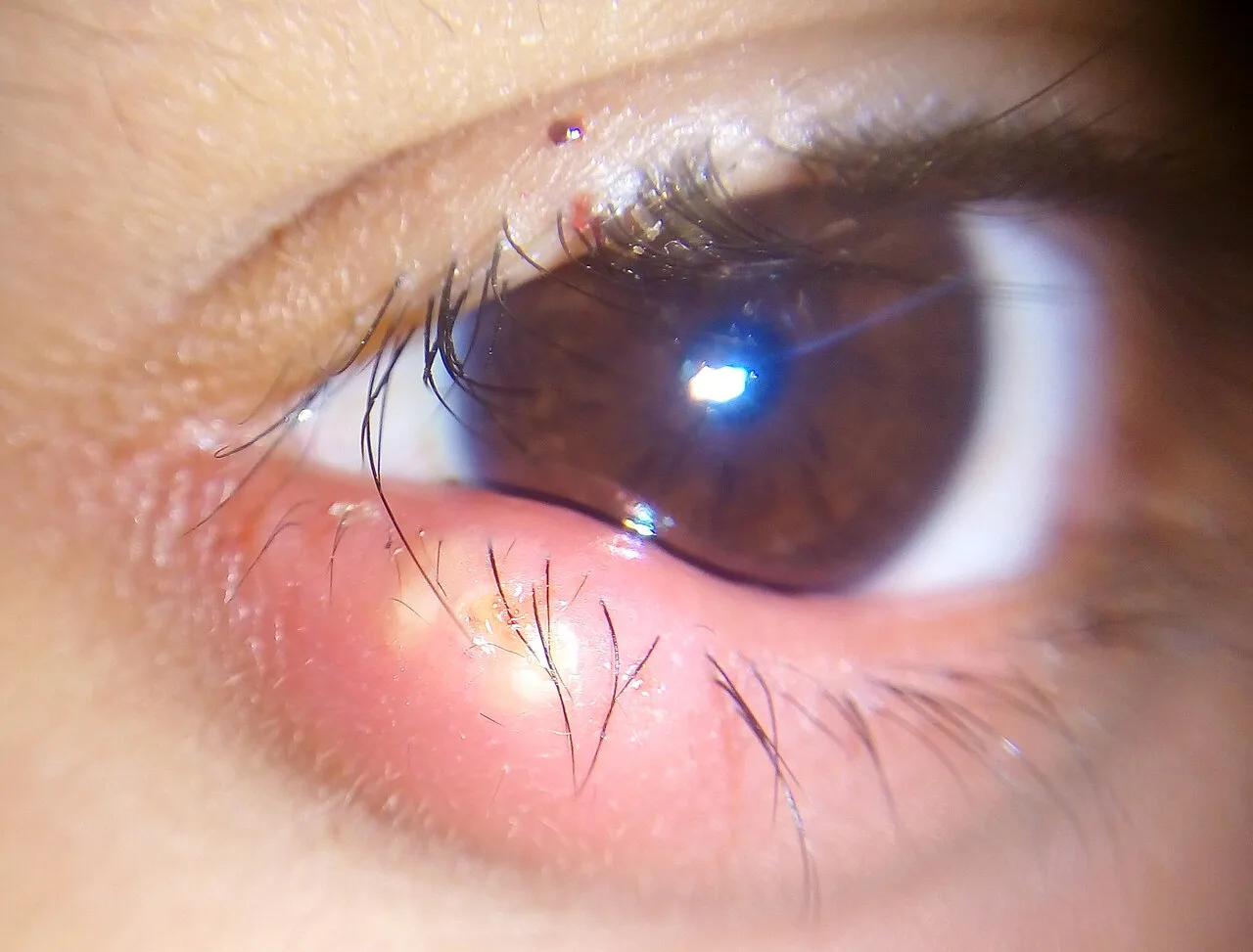
The lower lid carries the same meibomian glands and lash follicles as the upper, so it gets the same chalazia, styes and blepharitis, and it is the commoner site of basal cell carcinoma, because it takes more sun. Any lid lump that destroys the lashes, ulcerates or keeps returning after drainage is biopsied.
Examine it
Laxity of the lower lid turns it in (entropion) or out (ectropion).
Ask OphthoSpace about the lower tarsal plate
AI responses are for education and may contain errors. OphthoSpace is not for primary clinical diagnosis.