Visual atlas
See it before you are asked to name it
132 real images across 7 modality groups. Every one was opened on its Wikimedia Commons file page and checked for what it shows, its licence and whether anyone could be recognised. Tap an image to study it; the practice case behind it grades your localisation and diagnosis.
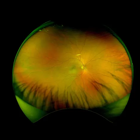 Image of the dayUltra-widefield image at a myopia reviewTry it blind
 12 images
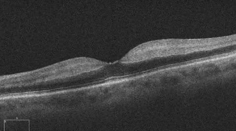 OCT maculaLabelled
Macular OCT hours after sudden visual loss
Same presentation: sudden painless loss of vision in one eye with a relative afferent pupillary defect; OCT of the macula.
advanced · retina
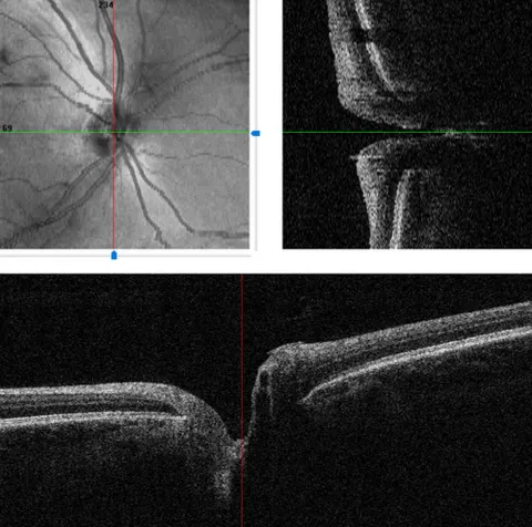 OCT optic nerveLabelled
Optic nerve head OCT with scan positions
Baseline disc imaging in a glaucoma suspect; the software shows the en face image and two B scans through the disc.
advanced · glaucoma
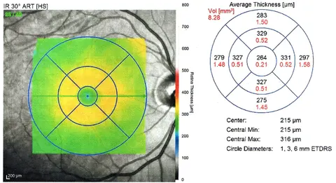 OCT maculaLabelled
Macular thickness map at baseline
A 50-year-old with diabetes and no retinopathy; baseline macular OCT thickness map of the right eye.
intermediate · retina
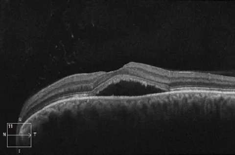 OCT macula
A grey smudge in the centre of vision in a stressed 38-year-old
A 38-year-old man in a stressful job, using a steroid nasal spray, with two weeks of a grey smudge in the centre of the left eye and objects looking smaller. Acuity 6/9.
intermediate · retina
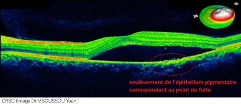 OCT maculaLabelled
The leak point and the fluid on a labelled OCT
A labelled OCT of a patient with central serous chorioretinopathy, with the pigment epithelial elevation at the leak point marked.
advanced · retina
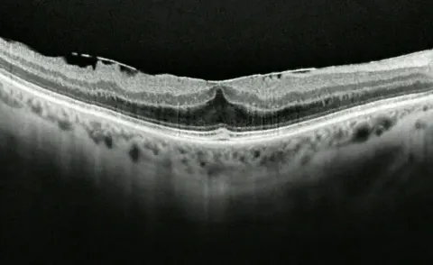 OCT macula
Straight lines look wavy, slowly, in one eye at 72
A 72-year-old notices over a year that straight lines look wavy in one eye and small print is harder. Acuity 6/12.
intermediate · retina
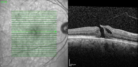 OCT macula
The scan pattern and the cut through the hole
A 70-year-old with months of central distortion and a small blank spot in the right eye. Acuity 6/36.
intermediate · retina
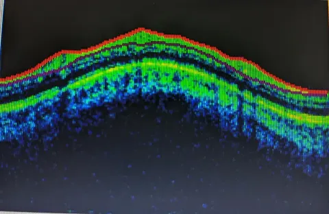 OCT macula
A normal macula on a colour-coded OCT
The right eye of a 53-year-old man with no symptoms, scanned at a routine examination.
foundational · comprehensive
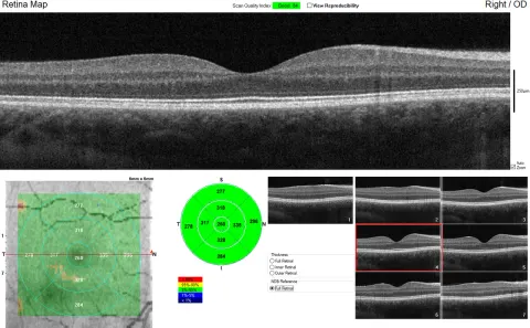 OCT maculaLabelled
The full macular OCT report of a healthy 24-year-old
The right eye of a healthy 24-year-old man, scanned to show what a normal report looks like.
foundational · comprehensive
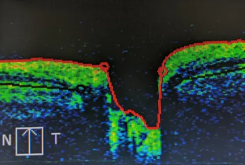 OCT optic nerve
A cut through the optic nerve head
An OCT cross-section through the optic disc and the retina on either side of it.
intermediate · glaucoma
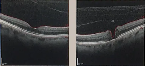 OCT macula
A small central blank spot and bent lines, over months
A 67-year-old woman with a few months of a small blank spot in the centre of the right eye and straight lines that bend. Acuity 6/24.
intermediate · retina
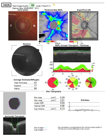 OCT optic nerveLabelled
The optic nerve head report of a right eye with glaucoma
The right eye of an 80-year-old man under treatment for glaucoma, imaged at review.
advanced · glaucoma