Interpret/6 pages
Read the images ophthalmology runs on.
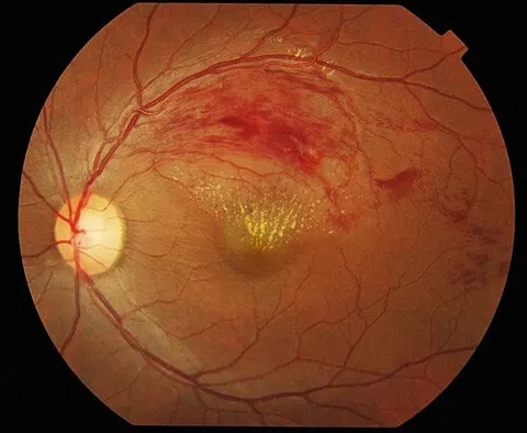
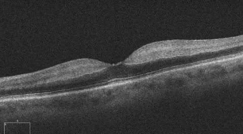
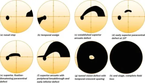
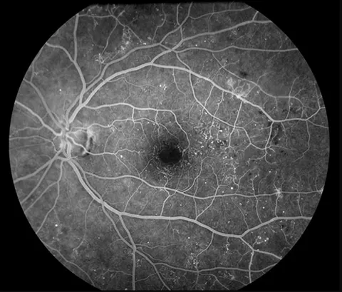
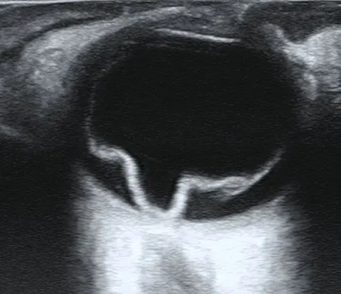
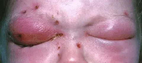
Each modality answers a different question. The fundus photograph shows the surface; OCT the layers beneath it; the field the function; angiography the flow; ultrasound the shape when there is no view. Every image here is real and the diagnosis waits until you commit.
0 of 6 explored