Learn / Glaucoma
Glaucoma
The disc, the pressure, the angle, the field.
Glaucoma is an optic neuropathy with a characteristic disc and field, for which intraocular pressure is the main treatable risk factor. It is not a pressure reading. Four things are read together: the disc, the pressure, the angle and the field.
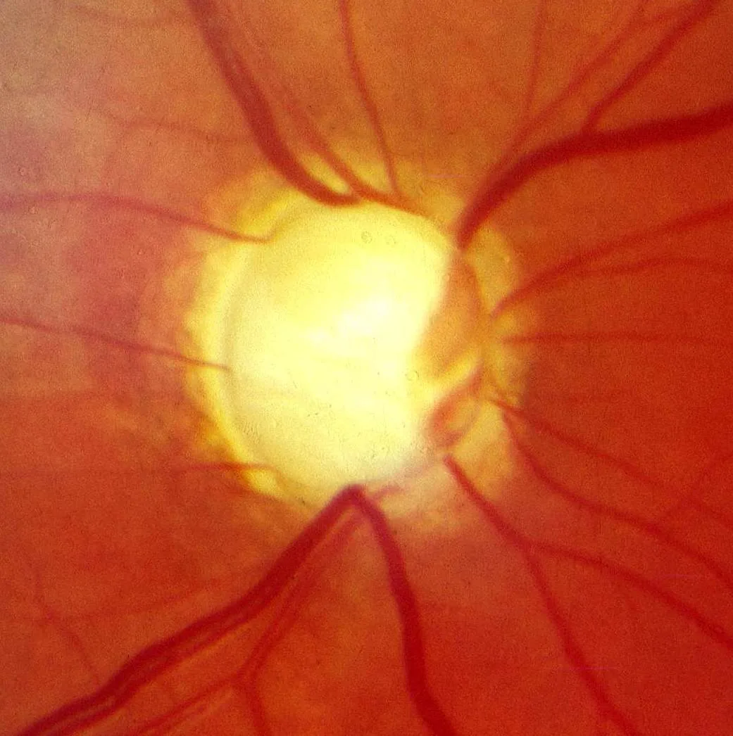
A disc found at a routine sight test, in a patient with no symptomsOpen in the atlas
Aqueous and pressure
Aqueous is made by the ciliary processes, flows through the pupil into the anterior chamber, and drains mostly through the trabecular meshwork at the angle into Schlemm's canal, with a fraction leaving by the uveoscleral route. Pressure is the balance of production and outflow; normal is roughly 10 to 21 mmHg, with a diurnal swing, and measured by applanation.
Pressure alone does not define glaucoma. Ocular hypertension is a high pressure with a normal nerve; normal-tension glaucoma is a glaucomatous nerve with a pressure in the normal range. The nerve decides.
Tonometry and what pressure means The ciliary body on the 3D eye
The glaucomatous disc
Ganglion cell axons die and the neuroretinal rim thins, so the cup enlarges and deepens. Read the rim, not the cup: the ISNT rule says the normal rim is thickest inferiorly, then superiorly, nasally, temporally; a rim that breaks the rule, a notch, a vertical cup larger than the horizontal, a splinter haemorrhage at the margin, nasal displacement and bayoneting of the vessels, and asymmetry of more than 0.2 between the eyes all point to glaucoma. OCT of the retinal nerve fibre layer measures the loss.
- A verified photograph of glaucomatous cupping is not yet in the atlas; the normal disc and its OCT are here for the baseline.
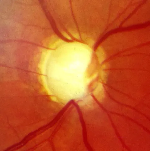
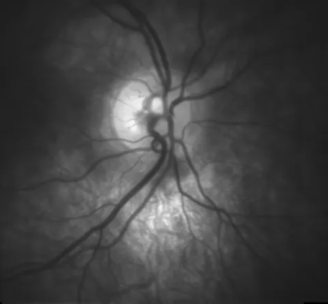
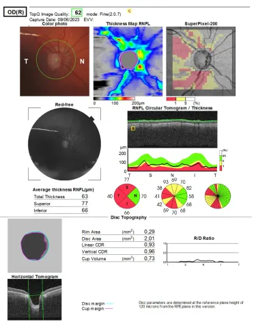
Primary open-angle glaucoma
The common form: an open angle with impaired outflow through the meshwork, raised or normal pressure, a slowly cupping disc and a field that goes from the periphery and the arcuate bundles inwards, painlessly and unnoticed until late. Risk rises with age, pressure, family history, African ancestry, myopia and steroid use. Treatment lowers the pressure: prostaglandin analogue drops first, then beta-blockers, carbonic anhydrase inhibitors, alpha agonists, laser trabeculoplasty, and surgery (trabeculectomy, drainage devices) when drops fail.
Acute angle closure
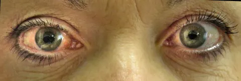
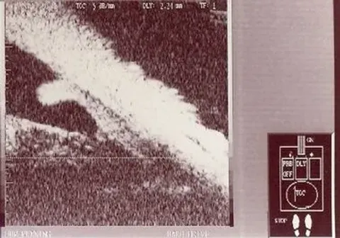
Emergent · nowIn a short, hypermetropic eye with a thick lens, the iris root can be pushed against the meshwork and shut the drain. When it shuts suddenly, the pressure climbs to 50 or 60 mmHg within hours: a severe aching eye and headache, nausea and vomiting, blurred vision with haloes around lights, a red eye with a cloudy cornea, a fixed mid-dilated pupil and a stone-hard globe. It happens in the evening, in dim light, sometimes after a dilating drop or an anticholinergic.
It is an emergency: pressure-lowering drops and intravenous acetazolamide, then laser peripheral iridotomy in both eyes once the cornea clears, because the fellow eye has the same anatomy.
The field
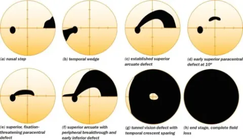
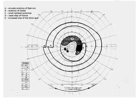
Glaucoma takes the arcuate nerve fibre bundles first, so the defects respect the horizontal midline: arcuate scotomas, a nasal step, then a temporal island and central vision last. The field is followed as a series; one printout is a photograph, the trend is the disease.
Go and look
Where this topic leads