Presentations / Diplopia
Diplopia
Cover one eye. Does it go?
Double vision that disappears when either eye is covered is binocular: the eyes are misaligned, and the question is which nerve or muscle. Double vision that stays with one eye open is monocular: an optical problem in that eye, never a nerve.
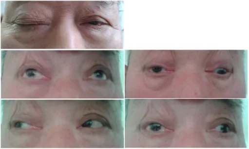
The nine positions of gaze with one nerve outOpen in the atlas
Monocular or binocular
Monocular diplopia (a ghost image that persists with the other eye shut) is astigmatism, an early cataract, a dry eye, a dislocated lens: refraction and a slit lamp. Binocular diplopia is the rest of this page.
Which direction, and where worst
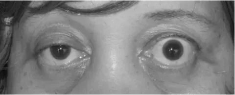
Horizontal diplopia worst looking to one side and at distance: the lateral rectus on that side, a sixth nerve palsy. Vertical or oblique diplopia worst looking down and in, with a head tilt: a fourth nerve palsy. A ptosis, an eye down and out and a big pupil: a third nerve palsy, and a call about the pupil. Diplopia that is worse at the end of the day with a variable ptosis: myasthenia. Restricted upgaze with lid retraction and proptosis: thyroid eye disease. Restricted upgaze after a blow, with a numb cheek: an orbital floor fracture trapping the inferior rectus. An adducting eye that lags behind with nystagmus of the other: an internuclear ophthalmoplegia, from the medial longitudinal fasciculus, and in a young adult, multiple sclerosis.
| Cause | Diplopia | Signs | The point |
|---|---|---|---|
| Sixth nerve palsy | Horizontal, worse to the affected side and at distance | Cannot abduct, esotropia | Vascular in the older diabetic; raised pressure or a tumour otherwise |
| Fourth nerve palsy | Vertical, worse down and in | Hypertropia, head tilt away | Trauma, congenital decompensation, microvascular |
| Third nerve palsy | Oblique, in every direction, if the lid is lifted | Ptosis, down and out, pupil | Painful with a big pupil: aneurysm, same day |
| Myasthenia gravis | Variable, worse late in the day | Fatigable ptosis, no pupil involvement | Antibodies, ice test, the rest of the muscles |
| Thyroid eye disease | Worst in upgaze | Lid retraction, proptosis, restricted inferior rectus | Thyroid function, the orbit on CT |
| Orbital floor fracture | Vertical, worst in upgaze | Enophthalmos, numb cheek, restricted elevation | CT; surgery if the muscle is trapped |
| Internuclear ophthalmoplegia | Horizontal on side gaze | Adduction lag, abducting nystagmus | MRI: demyelination in the young, stroke in the old |
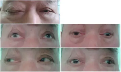
Examine it
Look for a head posture, a ptosis, a squint at rest and the corneal reflections. Cover test. Ductions and versions through the nine positions, asking where the images separate most. The pupils, always. The lids, for fatigue. The fundi, for papilloedema. And the rest of the cranial nerves, because a sixth nerve palsy with a fifth nerve deficit is a cavernous sinus, not a microvascular event.
Go and look
Where this topic leads