Presentations / Sudden vision loss
Sudden vision loss
Painless or painful, one eye or both, seconds or hours.
Sudden loss of vision is a vascular event, a detachment, a haemorrhage or an inflamed nerve until proven otherwise. The history sorts it before the examination does: how fast, how painful, one eye or both, and what the patient means by loss.
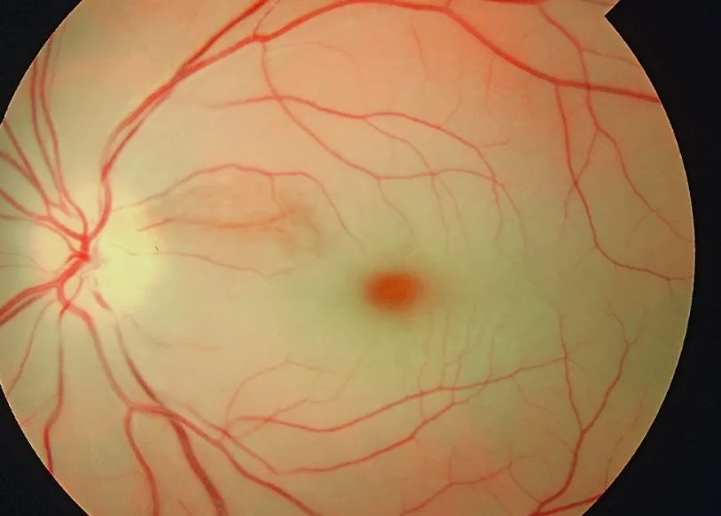
Vision gone in seconds, painless, an hour agoOpen in the atlas
Sort it by the history
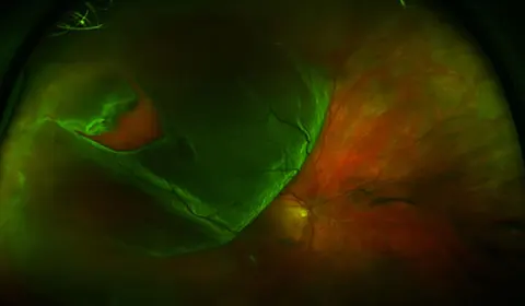
Seconds and painless: an arterial event (central retinal artery occlusion, or amaurosis fugax if it recovered within minutes). Hours and painless with a fundus full of blood: a vein occlusion. Floaters and flashes then a curtain: a detachment. A sudden shower of floaters and a red haze: vitreous haemorrhage. Days, with pain on eye movement, in a young adult: optic neuritis. On waking, painless, in someone over fifty, with an altitudinal field: ischaemic optic neuropathy, and the question of arteritis. Both eyes at once, or a hemianopia: the brain, and a stroke pathway.
| Cause | Speed | Pain | Fundus | Pupil | Urgency |
|---|---|---|---|---|---|
| Central retinal artery occlusion | Seconds | None | Pale retina, cherry red spot | RAPD | Emergent: source and arteritis |
| Amaurosis fugax | Seconds, recovers in minutes | None | Normal, or an embolus in a vessel | Normal after | Urgent: a stroke warning, carotid and heart |
| Central retinal vein occlusion | Hours | None | Haemorrhages in all quadrants, dilated veins, swollen disc | RAPD if ischaemic | Urgent |
| Retinal detachment | Hours to days, a curtain | None | Grey elevated retina | RAPD if large | Emergent while the macula is on |
| Vitreous haemorrhage | Minutes to hours, a red haze and floaters | None | No view, or blood in the vitreous | Normal | Urgent: find the cause (diabetes, a tear) |
| Optic neuritis | Days | On eye movement | Normal or swollen disc | RAPD | Soon: MRI |
| Arteritic ischaemic optic neuropathy | Hours, on waking | Headache, jaw claudication | Pale swollen disc | RAPD | Emergent: steroids on suspicion |
| Wet macular degeneration | Days, distortion | None | Subretinal fluid and haemorrhage at the macula | Normal | Urgent: anti-VEGF within days |
| Occipital stroke | Seconds to minutes, both eyes, a hemianopia | None or headache | Normal | Normal | Emergent: stroke pathway |
The examination that sorts it
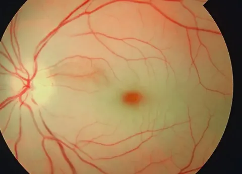
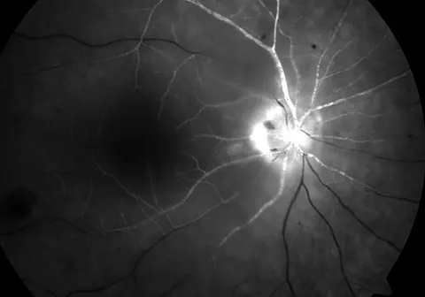
Acuity and the pinhole. The pupils, for an afferent defect, which means the retina or the nerve on that side is not sending. Confrontation fields, for an altitudinal defect (the nerve) or a hemianopia (the brain). The red reflex, lost in a vitreous haemorrhage. The fundus: a pale retina, blood, a grey elevation, a swollen or pale disc. And in anyone over fifty: the temporal arteries, the ESR and the CRP, today.
Pupil Lab: the RAPD Visual Field Lab: altitudinal and homonymous
The one that takes the other eye
Emergent · nowGiant cell arteritis. Any patient over fifty with sudden vision loss is asked about headache, scalp tenderness, jaw claudication, shoulder stiffness, weight loss and fever, and has an ESR and CRP before they leave. If the story fits, high-dose steroids start before the biopsy. The first eye is often lost; the second eye is what the treatment saves.
Go and look
Where this topic leads