Learn / Retina
Retina
Vascular disease, detachment, the macula.
The retina is the only place in the body where you look directly at blood vessels and neural tissue. Diabetes and hypertension write on it, veins and arteries block in it, the vitreous tears it, and the macula ages.
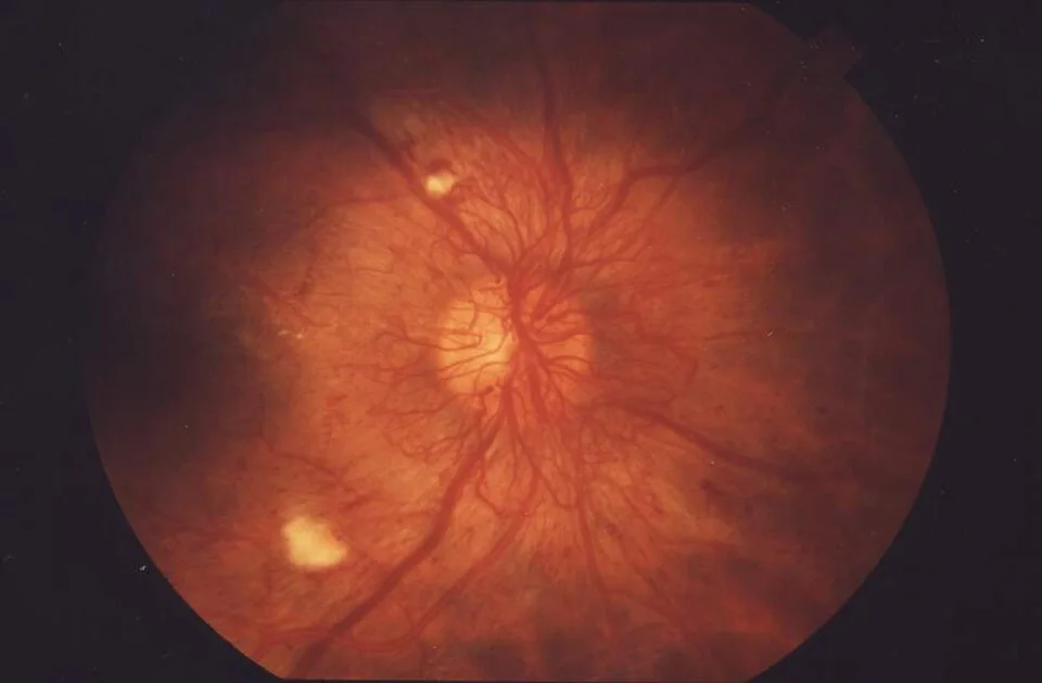
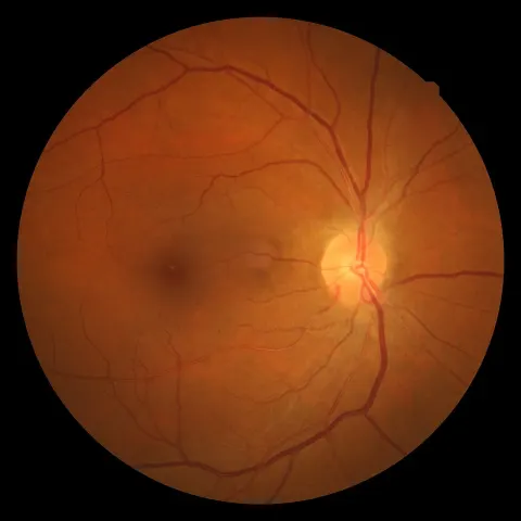
Floaters in a patient with long-standing type 1 diabetesOpen in the atlas
Diabetic retinopathy
Chronic hyperglycaemia damages capillary walls: pericytes die, microaneurysms bulge, vessels leak (haemorrhage, hard exudate, oedema) and close (cotton wool spots, venous beading, ischaemia). Ischaemic retina makes vascular endothelial growth factor, and VEGF grows new vessels on the disc (NVD) and elsewhere (NVE): proliferative retinopathy. The new vessels bleed into the vitreous and scar, and the scar pulls the retina off.
Vision is lost two ways: macular oedema at any stage (the commonest cause), and the complications of proliferative disease. Screening finds it before symptoms; control of glucose, pressure and lipids slows it; laser (panretinal for proliferative disease), anti-VEGF injections (for macular oedema and now for proliferative disease) and vitrectomy treat it.
| Stage | What you see | What it means |
|---|---|---|
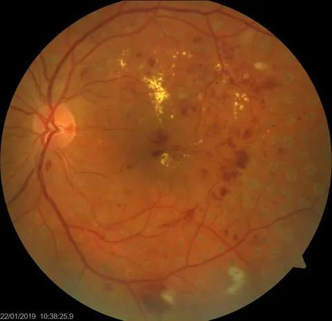
| Non-proliferative, mild to moderate | Microaneurysms, dot and blot haemorrhages, hard exudates | Leak; watch the macula |
| Non-proliferative, severe | Haemorrhages in four quadrants, venous beading, IRMA | Ischaemia; proliferation is coming |
| Proliferative | New vessels on the disc or elsewhere, preretinal or vitreous haemorrhage | Needs panretinal laser or anti-VEGF now |
| Diabetic maculopathy | Exudate and thickening at the macula, at any stage | Threatens central vision; OCT measures it |
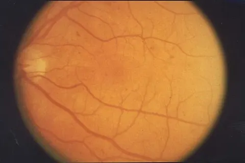
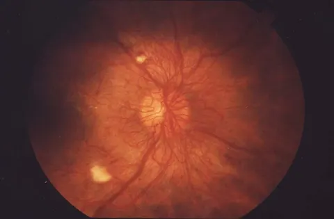
Compare non-proliferative with proliferative Diabetic macular oedema on OCT
Vein and artery occlusions
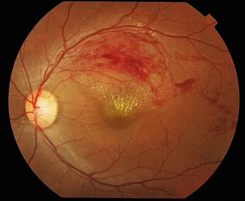
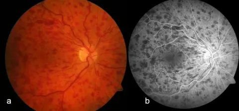
A retinal vein occlusion is a thrombosis at an arteriovenous crossing (branch) or at the lamina cribrosa (central): sudden painless blur, a fundus full of flame haemorrhages, dilated tortuous veins, cotton wool spots, and swelling. A branch occlusion keeps to one sector; a central one covers the whole fundus (blood and thunder). Vision is lost to macular oedema, treated with anti-VEGF, and to neovascular glaucoma in ischaemic cases.
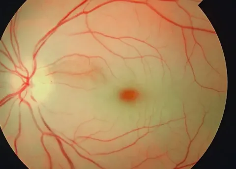
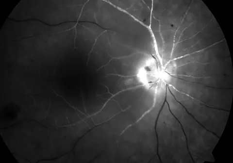
A central retinal artery occlusion is a stroke of the eye: an embolus from the carotid or the heart, or giant cell arteritis. Painless loss of vision in seconds to hand movements, an afferent pupillary defect, a pale oedematous retina with a cherry red spot at the fovea, and on OCT a bright, thickened inner retina. There is no proven treatment once hours have passed; the work is finding the source, and excluding arteritis in anyone over fifty.
Retinal tears and detachment
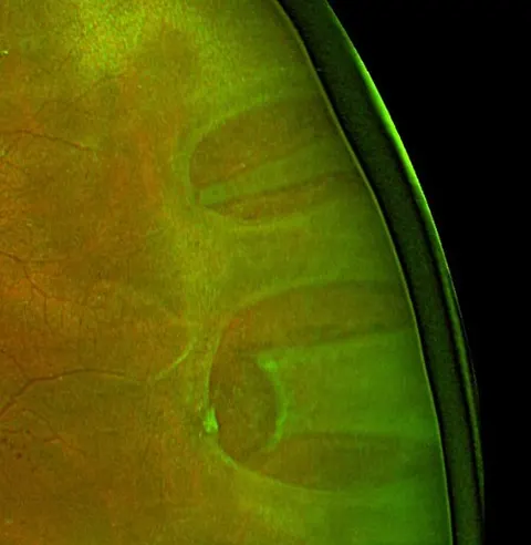
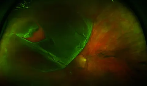
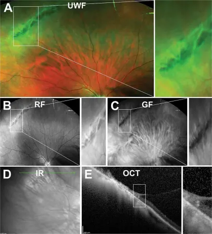
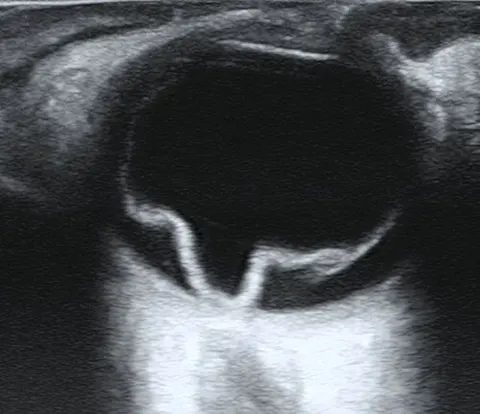
The vitreous liquefies with age and pulls away from the retina (posterior vitreous detachment): flashes from traction and floaters from the collapsing gel. Where it is stuck, it tears the retina, and fluid through the tear lifts the retina off the pigment epithelium: a rhegmatogenous detachment. A shadow or curtain spreads from the periphery towards the centre.
The retina looks grey, elevated and corrugated with darker vessels; the tear is at the edge of the detachment, and every tear must be found. If the macula is still on, surgery is within a day or so; once it is off, the visual result is worse and the timing less critical. A tear without a detachment is lasered to wall it off.
The macula and age-related macular degeneration
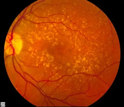
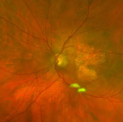
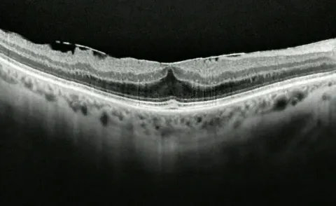
Drusen, yellow deposits under the pigment epithelium, are the substrate of age-related macular degeneration. Dry AMD is drusen and atrophy: slow loss of central vision. Wet (neovascular) AMD is a choroidal neovascular membrane growing through Bruch's membrane and leaking: distortion (straight lines bend) and rapid central loss, with subretinal fluid, haemorrhage and a grey membrane at the macula. OCT shows the fluid; anti-VEGF injections, repeated, keep it dry.
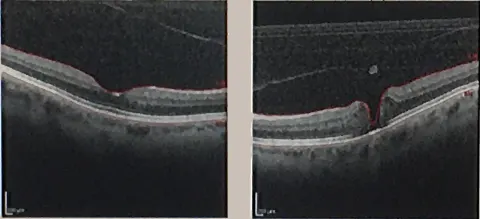
Central serous chorioretinopathy is a smooth dome of subretinal fluid in a younger, often stressed, patient, usually resolving; macular hole and epiretinal membrane are the vitreous pulling at the fovea. All are read on OCT.
- Verified fundus photographs of drusen, wet AMD, macular hole and epiretinal membrane are not yet in the atlas; the OCT Lab carries real scans of drusen and choroidal neovascularisation.
The choroid
The vascular coat beneath the retina, supplying the outer retina and the photoreceptors. Choroidal neovascularisation is the lesion of wet AMD; posterior uveitis and choroiditis (sarcoid, toxoplasmosis, tuberculosis) inflame it; choroidal melanoma is the commonest primary intraocular tumour in adults, a pigmented dome under the retina found at a routine examination.
Hypertensive retinopathy
Chronic hypertension narrows the arterioles, thickens their walls (copper and silver wiring) and pinches the veins where they cross (arteriovenous nicking). Accelerated hypertension adds flame haemorrhages, cotton wool spots and hard exudate, and at its worst a swollen disc: malignant hypertension, a medical emergency whose eye signs are the reason to look.
- A verified photograph of hypertensive retinopathy is not yet in the atlas.
Go and look
Where this topic leads