Interpret / Fluorescein angiography
Fluorescein angiography
Leak, block, and where the blood does not go.
Fluorescein injected into an arm vein reaches the eye in ten to fifteen seconds and is photographed through a blue exciter and a yellow barrier filter as it fills the choroid, the arteries, the capillaries and the veins. It shows what a photograph cannot: which vessels leak, which are blocked, and where the capillaries have closed.
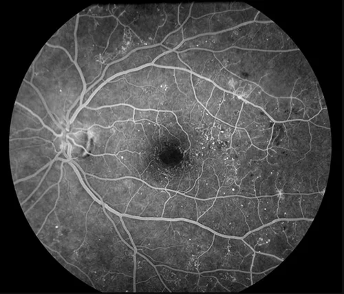
Angiogram for unexplained central visual loss in diabetesOpen in the atlas
The phases
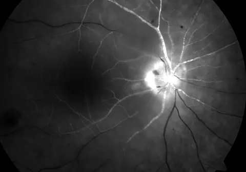
Choroidal flush at about ten seconds, patchy then confluent. Arterial phase a second later. Arteriovenous phase: the arteries full, the veins filling in laminar streams along their walls. Venous phase by about 30 seconds. Late phase at five to ten minutes, when normal vessels have emptied and anything that leaked or stained still glows. A delay in the arterial filling, or an arm-to-retina time beyond about 15 seconds, means the artery is obstructed.
Hyper and hypo
Every abnormality is either too much fluorescence or too little, and each has a short list.
| Pattern | Mechanism | Examples |
|---|---|---|
| Hyperfluorescence: leak | Dye escapes an abnormal vessel and spreads over time | Diabetic macular oedema, new vessels, cystoid macular oedema (a petaloid pattern), wet AMD |
| Hyperfluorescence: pooling | Dye collects in a space | A pigment epithelial detachment, central serous chorioretinopathy (a smokestack) |
| Hyperfluorescence: staining | Tissue holds dye late | Drusen, scars, the disc |
| Hyperfluorescence: window defect | Thin RPE lets the choroidal glow through, without leak | Atrophy, old laser scars |
| Hypofluorescence: blocking | Something in front hides the glow | Haemorrhage, pigment, exudate |
| Hypofluorescence: filling defect | Blood does not arrive | Capillary non-perfusion in diabetes and vein occlusion, an artery occlusion |
When it is used
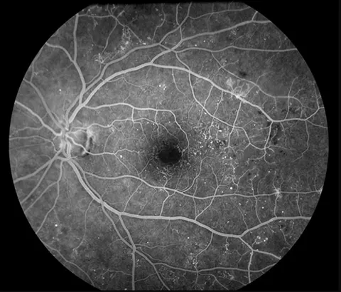
To map non-perfusion and find new vessels in diabetic retinopathy and vein occlusion, deciding where laser goes; to confirm and classify a choroidal neovascular membrane before treatment; to explain a macular oedema; to time an arterial occlusion. OCT and OCT angiography have taken over much of this without a needle, but the dye study still shows leakage, which OCT angiography cannot. Indocyanine green angiography images the choroid through pigment and blood, for polypoidal disease and choroidal tumours.
Go and look
Where this topic leads