Interpret / OCT Lab
The retina in cross section
Optical coherence tomography slices the macula at the resolution of histology. Learn the layers, then read real scans the same way every time: the foveal contour, the fluid and which compartment it is in, what the pigment epithelium is doing, and only then the impression.
Read the scan
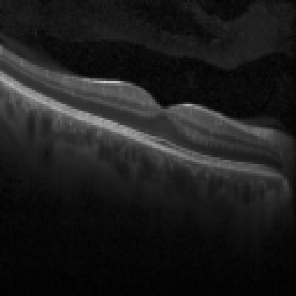
Kermany DS, Zhang K, Goldbaum M. Labeled Optical Coherence Tomography (OCT) and Chest X-Ray Images for Classification. Mendeley Data, V2 (2018). CC BY 4.0. Cut from the dataset's contact sheets; research and educational use, not clinical diagnosis. Dataset. Greyscale unaltered; brightness and contrast are the viewer's own controls.
Read it in order
1. Foveal contour
2. Fluid
3. The RPE
4. Impression
The layers, vitreous to choroid
Alternating bright and dark bands: plexiform layers and the nerve fibre layer are bright, the nuclear layers dark, the RPE the brightest line of all.
- 01
Internal limiting membrane ILM
The bright inner surface of the retina against the black vitreous. Epiretinal membranes lie on it.
- 02
Retinal nerve fibre layer RNFL
Bright, thickest near the disc, absent at the fovea. Measured around the disc for glaucoma.
- 03
Ganglion cell layer GCL
Darker, thick around the fovea and thin at it; lost early in glaucoma and optic neuropathy.
- 04
Inner plexiform layer IPL
Bright: synapses between bipolar and ganglion cells.
- 05
Inner nuclear layer INL
Dark: the bipolar, horizontal and amacrine cell bodies. Cystoid spaces of macular oedema sit here and in the outer plexiform layer.
- 06
Outer plexiform layer OPL
Bright: photoreceptor synapses. Hard exudate collects here as bright dots with shadows.
- 07
Outer nuclear layer ONL
Dark and thick at the fovea: the photoreceptor cell bodies, all cones at the centre.
- 08
External limiting membrane ELM
A thin bright line; its integrity predicts visual recovery.
- 09
Ellipsoid zone EZ
A bright band of photoreceptor inner segments; disruption here correlates with reduced vision.
- 10
Retinal pigment epithelium RPE
The brightest outer band, with Bruch's membrane beneath. Drusen bump it; a pigment epithelial detachment lifts it; atrophy loses it and lets light through to the choroid.
- 11
Choroid Choroid
Vessels as dark ovals below the RPE, on newer scans down to the sclera.
Normal beside abnormal
More OCT in the atlas
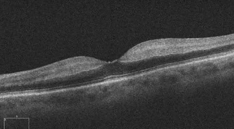
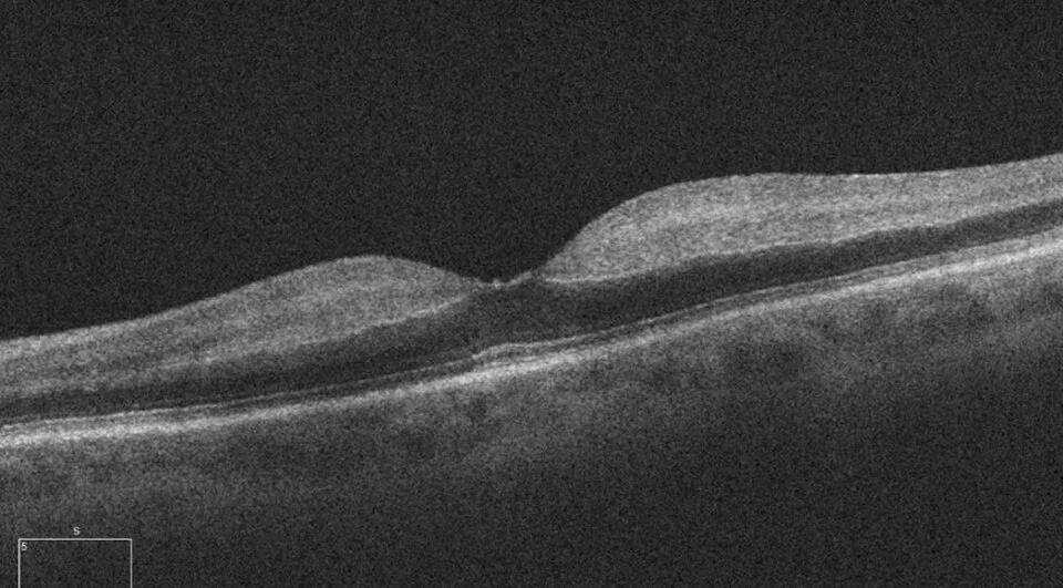
Macular OCT hours after sudden visual loss
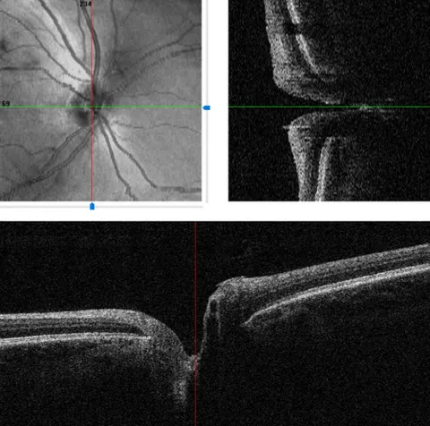
Optic nerve head OCT with scan positions
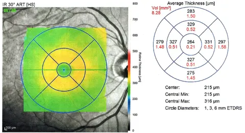
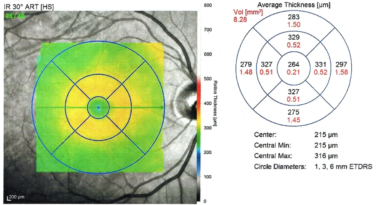
Macular thickness map at baseline
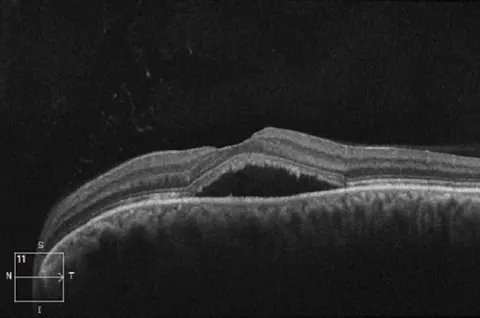
A grey smudge in the centre of vision in a stressed 38-year-old
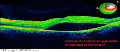
The leak point and the fluid on a labelled OCT
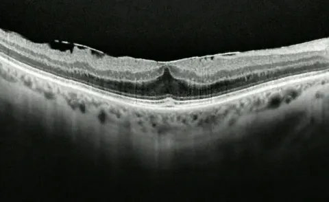
Straight lines look wavy, slowly, in one eye at 72
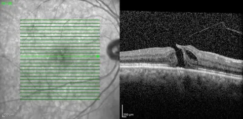
The scan pattern and the cut through the hole
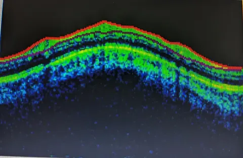
A normal macula on a colour-coded OCT
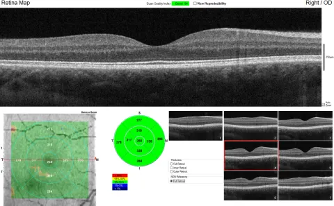
The full macular OCT report of a healthy 24-year-old
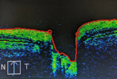
A cut through the optic nerve head
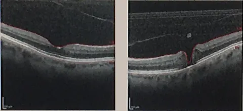
A small central blank spot and bent lines, over months
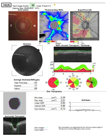
The optic nerve head report of a right eye with glaucoma
Taught here, not yet illustrated
These patterns matter and are described; a verified, openly licensed scan of each has not yet been found, so no image is shown rather than an invented one.
- Macular hole
- A full-thickness defect at the fovea with cystic edges; the contour is a hole rather than a pit. Vision loss is central and distortion is the complaint.
- Epiretinal membrane
- A bright line on the inner surface wrinkling the retina beneath, with a distorted or lost foveal pit. Metamorphopsia is the symptom.
- Central serous chorioretinopathy
- A smooth dome of subretinal fluid with a flat or slightly bumped RPE and a thick choroid, in a younger patient, often a stressed one.
- Retinal detachment on OCT
- The neurosensory retina lifted away from the RPE by a dark space, at the edge of the scan or across it.
Correlate every OCT with the fundus photograph: they answer different questions. Read the fundus