Interpret / Ocular ultrasound
Ocular ultrasound
The shape of the eye when there is no view.
B-scan ultrasound draws a cross section of the globe from a probe on the closed lid, through a cataract, a vitreous haemorrhage or a corneal scar that has stopped every other view. It answers a few questions well: is the retina attached, is there a mass, is there a foreign body.
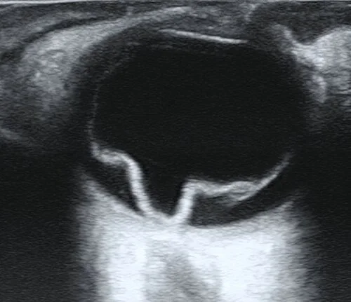
Ultrasound when the fundus cannot be seenOpen in the atlas
The image
The probe on the closed lid, with gel, at about 10 MHz. The cornea and lens at the top of the screen, the black vitreous, and the bright curve of the retina, choroid and sclera at the back with the optic nerve's dark shadow behind. Gain is turned down to make the vitreous black and up to bring out faint echoes. The eye is moved and the probe angled to look at every meridian.
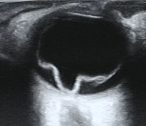
What it shows
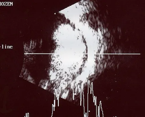
A retinal detachment is a bright, thick, mobile membrane attached at the disc and the ora serrata, folding with eye movement. A posterior vitreous detachment is a thinner, more mobile membrane not attached at the disc. Vitreous haemorrhage is diffuse low echoes that swirl. A choroidal melanoma is a dome or collar-stud mass with low internal reflectivity and a hollow behind it; a retinoblastoma is a mass with bright calcium that casts a shadow. An intraocular foreign body is a bright echo with shadowing and reverberation. The optic nerve sheath diameter can be measured on a transorbital view, a bedside marker of raised intracranial pressure.
At the bedside
Point-of-care ultrasound in the emergency department, on a closed eye with plenty of gel and no pressure, finds a detachment, a vitreous haemorrhage, a lens dislocation or a foreign body in minutes. It is never used on a suspected open globe: pressure on the lid pushes on the wound.
Go and look
Where this topic leads