Presentations / Flashes and floaters
Flashes and floaters
The vitreous letting go, and whether it took the retina with it.
New floaters and flashes are a posterior vitreous detachment until the periphery has been examined through a dilated pupil. Most are harmless. One in ten has a retinal tear, and a tear becomes a detachment.
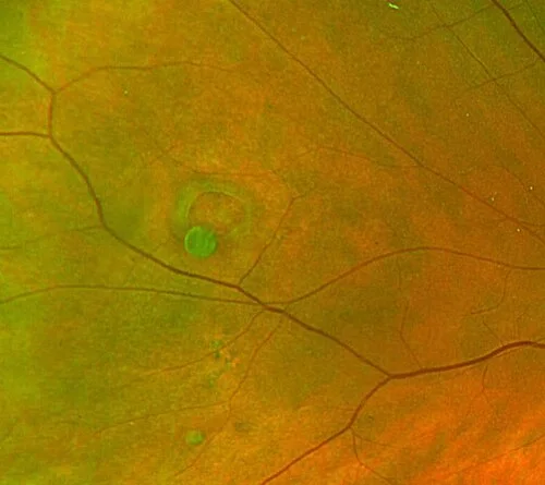
An incidental finding at a dilated examinationOpen in the atlas
What is happening
The vitreous gel liquefies with age and pulls away from the retina. The collapsing gel casts shadows: floaters, often one large ring where it left the disc. Where it tugs on the retina it fires it: flashes, brief arcs of light in the temporal field, in the dark. If it tugs hard enough where it is stuck, it tears the retina, and fluid through the tear lifts the retina off.
The worrying features
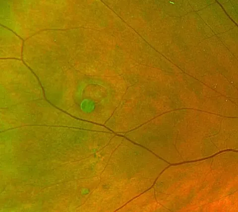
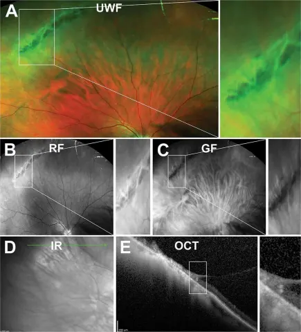
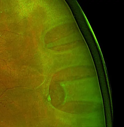
A shower of many new floaters (a tear has bled), a curtain or shadow in any part of the vision (a detachment has started), reduced acuity (the macula is involved), high myopia, recent cataract surgery, or a previous detachment in either eye. Pigment cells in the anterior vitreous (tobacco dust) at the slit lamp mean a tear until the periphery proves otherwise.
| Cause | The story | The finding | Urgency |
|---|---|---|---|
| Posterior vitreous detachment | A few floaters, flashes in the dark | A Weiss ring, no tear on a dilated examination | Soon: examined within days, and again if symptoms change |
| Retinal tear | A shower of floaters, flashes | A horseshoe tear at the periphery, pigment in the vitreous | Urgent: laser within a day or two |
| Retinal detachment | A curtain, a shadow, then central loss | Grey elevated retina | Emergent while the macula is on |
| Vitreous haemorrhage | A red haze, many floaters | No red reflex, or blood in the gel | Urgent: find the tear or the diabetic new vessels |
| Migraine aura | Shimmering zigzags in both eyes for twenty minutes, then a headache | Normal eye | Routine |
Examine it
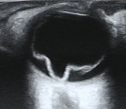
Acuity, then the pupils, then dilate. The slit lamp for pigment in the anterior vitreous. The peripheral retina with the indirect ophthalmoscope and indentation, or a widefield photograph, to find every tear. A B-scan ultrasound when a haemorrhage blocks the view.
Go and look
Where this topic leads