Anterior segment · 1 of 24
What is the cornea?
The cornea is the clear front window of the eye: a dome of transparent tissue about half a millimetre thick and eleven to twelve millimetres across, continuous with the white sclera at the limbus. It has no blood vessels, which is why it is clear, and it is the most densely innervated tissue in the body, which is why a scratch on it hurts so much. It provides about two thirds of the eye's focusing power.
- Thickness
- About 0.5 mm at the centre, thicker at the edge
- Diameter
- 11 to 12 mm
- Power
- About 43 dioptres, two thirds of the eye's total
- Nerve supply
- Long ciliary nerves, from the ophthalmic division of the trigeminal nerve
- Nutrition
- Tears in front, aqueous behind, limbal vessels at the edge; no vessels of its own
Five layers, front to back
The cornea is built in five layers, and each one has its own diseases. From the tear film inwards: the epithelium, Bowman's layer, the stroma, Descemet's membrane and the endothelium.
The epithelium is five or six cells thick and renews itself every week from stem cells at the limbus. It is the layer an abrasion removes and the layer fluorescein stains when it is missing. Bowman's layer is a tough acellular sheet beneath it that does not regenerate: an injury through Bowman's layer leaves a scar. The stroma is ninety per cent of the thickness, collagen lamellae laid down so regularly that light passes without scattering; oedema or infiltrate disturbs that order and the cornea turns hazy. Descemet's membrane is the basement membrane of the endothelium, and the endothelium is a single layer of cells that pumps water out of the stroma all day to keep it clear. Endothelial cells do not divide, so their number only falls with age, surgery and disease.
| Layer | What it is | What goes wrong |
|---|---|---|
| Epithelium | Five or six cell layers, renewed weekly from the limbus | Abrasion, dendritic ulcer, punctate keratopathy, recurrent erosion |
| Bowman's layer | Acellular collagen sheet, does not regenerate | Scarring after deep injury, band keratopathy in front of it |
| Stroma | Regular collagen lamellae, 90 per cent of the thickness | Infiltrate and ulcer, scarring, keratoconus, oedema |
| Descemet's membrane | The endothelium's basement membrane | Breaks in keratoconus and birth trauma, folds when the cornea swells |
| Endothelium | A single pumping cell layer that never divides | Fuchs dystrophy, keratic precipitates, decompensation after surgery |
What it does
The cornea does two things: it lets light in and it bends it. Its clarity comes from having no vessels, an orderly stroma and an endothelium that keeps the tissue slightly dehydrated. Its power comes from its curvature and the jump in refractive index from air to tissue, which is why the same curve bends light far less under water and why a swimmer without goggles sees a blur.
The tear film sits on top and is optically part of the cornea: a dry, irregular surface scatters light and drops acuity even when the cornea beneath is healthy. That is why blinking briefly sharpens the vision of a dry eye, and why a pinhole cannot fully correct it.
How to examine it
At the slit lamp, start with diffuse light at low magnification to see the whole cornea, then narrow the beam to a slit and turn the illumination arm to about forty five degrees. The slit now cuts an optical section through the cornea: a bright curved band whose thickness you can read, with the epithelium at its front edge and the endothelium at its back. Thickening means oedema; a break in the section means a defect; an infiltrate sits inside the band at a depth you can name.
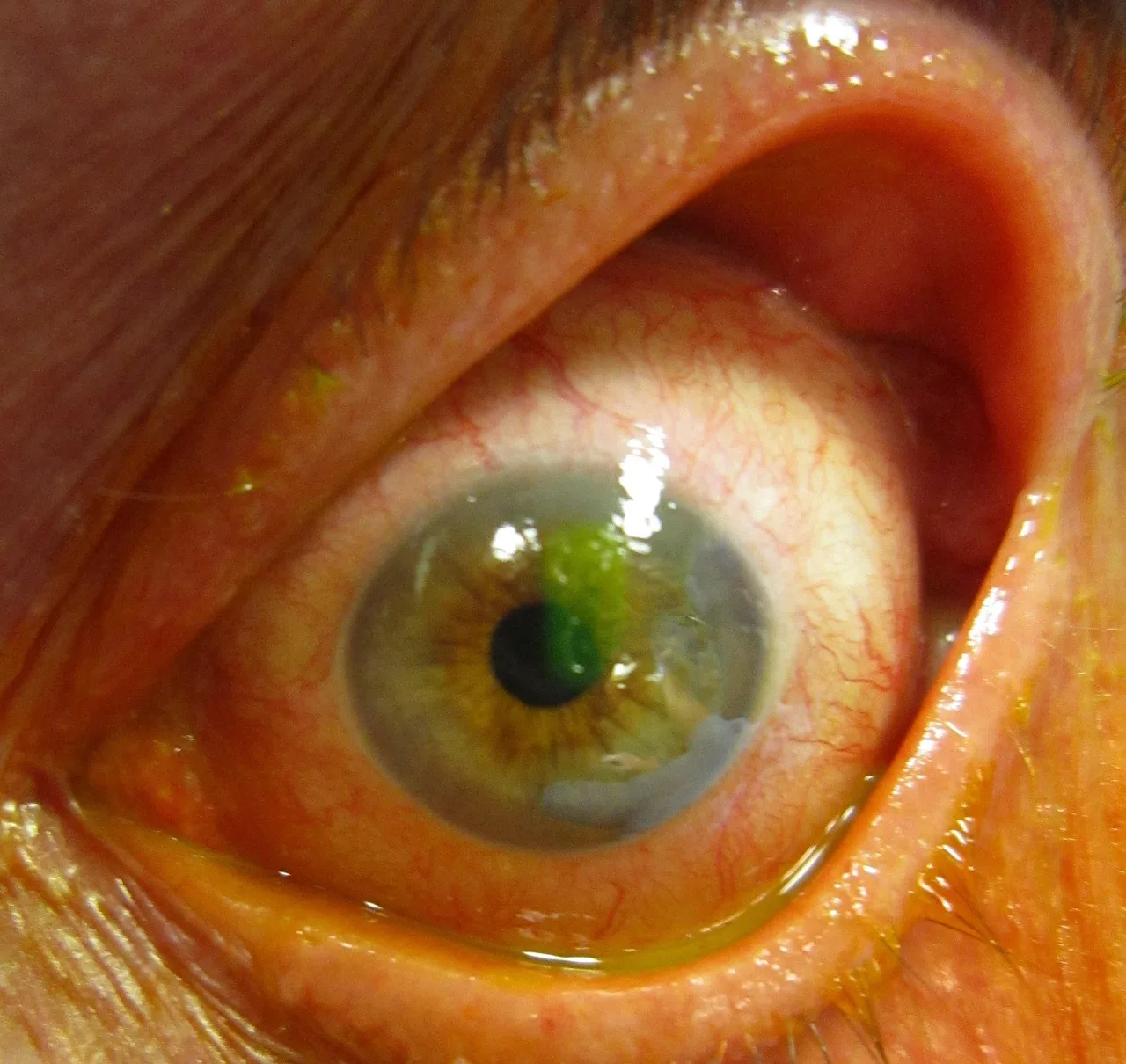
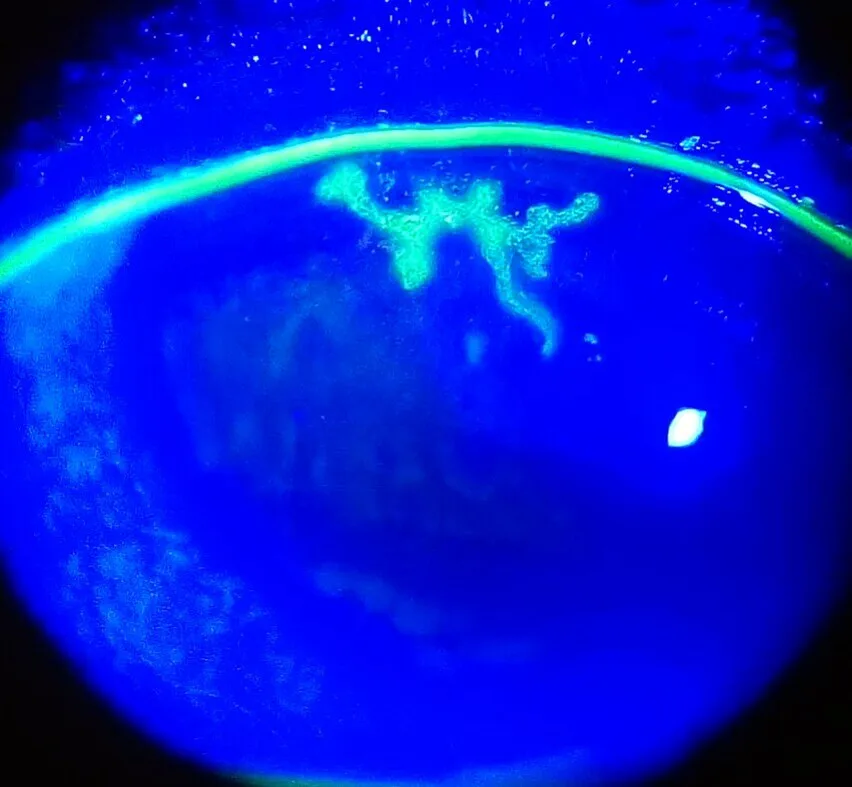
Then put a drop of fluorescein in and switch to cobalt blue light. Where epithelium is missing the dye pools and glows green: an abrasion is a sharp-edged patch, a dendritic ulcer a branching line with terminal bulbs, dry eye a scatter of fine dots. Check corneal sensation with a wisp of cotton before you put any drops in: a numb cornea points to herpes, and it is a sign you cannot recover once anaesthetic is on the eye.
What goes wrong
Corneal disease presents with pain, photophobia, watering and blur, in an eye that is red around the limbus rather than in the fornices. The questions that sort it out are whether the epithelium is intact, whether there is an infiltrate in the stroma, and whether the patient wears contact lenses.
An abrasion is a surface loss without infiltrate and heals in a day or two. A bacterial keratitis is an infiltrate with an overlying epithelial defect, usually in a contact lens wearer, and it is an emergency because it can perforate within days. Herpes simplex makes a dendrite, a branching ulcer that must not be treated with steroid. Keratoconus thins and steepens the cornea into a cone, blurring vision through irregular astigmatism in a young patient. Fuchs dystrophy is an endothelium that fails with age, so the cornea swells and vision is worst on waking.
- Contact lens wearer with a painful red eye: bacterial keratitis until proved otherwise; same-day review, no patching, no steroid.
- A dendrite means herpes simplex; steroid makes it worse, aciclovir makes it better.
- A white opacity on the cornea that stains is an ulcer; one that does not stain is a scar or a healed process.
- Hazy cornea, fixed mid-dilated pupil and a rock-hard eye is acute angle closure, not corneal disease.
Signs worth knowing
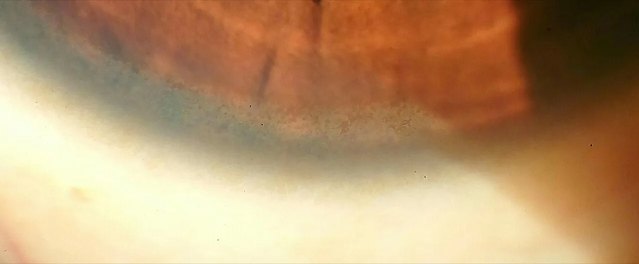
Arcus senilis is a grey ring at the corneal edge from lipid in the peripheral stroma, common and harmless after sixty and worth a lipid check before forty. A Kayser-Fleischer ring is copper in Descemet's membrane at the limbus and means Wilson disease. Band keratopathy is calcium in Bowman's layer, a horizontal grey band across the exposed cornea in chronic uveitis and hypercalcaemia.
Examine it
- Section the cornea in the slit lamp lab
- The cornea in the anterior segment topic
- Keratitis as an emergency
Abrasions, ulcers and dendrites live here; fluorescein and cobalt blue light show them.
Ask OphthoSpace about the cornea
AI responses are for education and may contain errors. OphthoSpace is not for primary clinical diagnosis.