Anterior segment · 2 of 24
What is the anterior chamber?
The anterior chamber is the space between the back of the cornea and the front of the iris, filled with clear aqueous humour. It is about three millimetres deep at the centre and shallows towards the edge, where cornea and iris meet at the angle. Because it is transparent and lit by the slit lamp, it is the one place in the body where you can watch inflammation happen: single cells drift through the beam like dust in sunlight.
- Depth
- About 3 mm at the centre in a normal adult eye, shallower with age and in hypermetropia
- Volume
- About a quarter of a millilitre of aqueous
- Aqueous turnover
- Made by the ciliary body at about 2 to 3 microlitres a minute, replaced every hundred minutes or so
- The angle
- Where iris root, ciliary body and cornea meet; the trabecular meshwork drains here into Schlemm's canal
The aqueous and where it goes
Aqueous humour is made by the ciliary processes behind the iris, in the posterior chamber. It flows forward through the pupil into the anterior chamber, and leaves at the angle through the trabecular meshwork into Schlemm's canal and the episcleral veins. A smaller share leaves through the ciliary body face, the uveoscleral route, which is the path prostaglandin drops open up.
Intraocular pressure is the balance of production and outflow. Block the meshwork and pressure rises slowly, the story of open angle glaucoma. Block the pupil so aqueous cannot reach the front, and the iris bows forward and closes the angle: pressure rises in hours, the story of acute angle closure.
How to examine it
The anterior chamber is examined by geometry. Narrow the slit to a short bright beam, about one by three millimetres, turn the illumination arm well to the side, and focus into the space between the corneal section and the iris. Against the dark pupil, the beam through healthy aqueous is invisible. If the beam is visible as a faint smoky path, that is flare: protein leaked from inflamed vessels. Bright specks drifting through the beam are cells, and they are counted by the number seen in that one by one millimetre field.
Depth is judged at the edge. Shine a narrow slit at the limbus with the arm at sixty degrees and compare the gap between cornea and iris with the thickness of the cornea itself: a gap of a quarter of the corneal thickness or less is a narrow angle at risk of closing. A torch held at the temporal limbus does the same job roughly: if the nasal iris falls into shadow, the chamber is shallow.
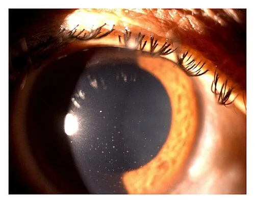
- 1Keratic precipitatesClumps of inflammatory cells stuck to the back of the cornea, settling inferiorly in a triangle (Arlt's triangle) as the aqueous convects.
- 2Finer precipitatesSmaller, dust-like deposits higher up. Fine KPs suggest non-granulomatous uveitis; large greasy 'mutton fat' KPs suggest granulomatous disease.
- 3Corneal light reflexThe lamp's reflection on the tear film. Normal, and a landmark for focusing on the front surface.
- 4IrisThe dilated pupil leaves a crescent of iris at the edge. Look at its margin for posterior synechiae.
What settles in it
Because the chamber is a fluid-filled space, what enters it settles by gravity, and the level it settles to is the diagnosis. Blood settles as a hyphaema, a red level at the bottom after blunt trauma. Pus settles as a hypopyon, a white level, and means severe inflammation: a bacterial corneal ulcer, endophthalmitis after surgery, or a violent uveitis. Both are examined with the patient upright, because the level only forms when the eye is still.
Cells and flare without a level are anterior uveitis: a painful, photophobic red eye with a small pupil, often in a young adult, and often with HLA-B27 disease elsewhere. Untreated, the inflamed iris sticks to the lens as posterior synechiae and the pupil becomes irregular.
| Finding | What it is | What it means |
|---|---|---|
| Cells | Single bright points drifting through the beam | Active inflammation; graded 0 to 4+ by the count in the beam |
| Flare | The beam visible as a smoky path | Protein in the aqueous; the blood-aqueous barrier is broken |
| Hypopyon | A white layered level | Severe inflammation or infection; a corneal ulcer or endophthalmitis until excluded |
| Hyphaema | A red layered level | Bleeding from the iris or angle after trauma; check pressure, rest upright, look for a rebleed |
| Shallow chamber | Iris close to the cornea at the edge | An occludable angle; do not dilate; acute closure if the pressure is high |
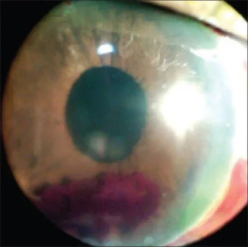
- 1Hyphaema, layeredBlood settled to the bottom of the anterior chamber with a horizontal fluid level, in front of the iris and behind the cornea.
- 2Fluid levelThe flat upper edge is the tell: blood layers by gravity, so the eye must be examined upright and left still.
- 3PupilRound and central. A peaked or torn pupil after trauma warns of an open globe or iris injury.
- 4Corneal light reflexSharp and undistorted: the cornea is clear, with no laceration under it.
The angle
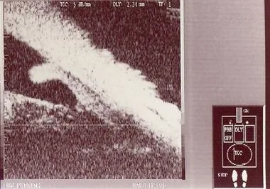
The angle cannot be seen directly, because light from it is reflected back into the eye at the cornea. Gonioscopy puts a contact lens with a mirror on the eye to see it: the structures from front to back are Schwalbe's line, the trabecular meshwork, the scleral spur and the ciliary body band. The more of them you can see, the more open the angle. Ultrasound biomicroscopy shows the same anatomy in cross section.
Examine it
- Find cells and a hyphaema in the slit lamp lab
- Cells, flare and hypopyon in the anterior segment topic
- Aqueous physiology and pressure
- Gonioscopy
Cells and flare mean uveitis; a hypopyon or hyphaema settles here; a shallow chamber warns of angle closure.
Ask OphthoSpace about the anterior chamber
AI responses are for education and may contain errors. OphthoSpace is not for primary clinical diagnosis.