Anterior segment · 3 of 24
What is the iris?
The iris is the coloured diaphragm of the eye, a thin disc of pigmented stroma and muscle with the pupil at its centre. It divides the anterior chamber from the posterior chamber, rests on the front of the lens, and changes the size of the pupil to control how much light reaches the retina. Its colour is only how much pigment sits in its front layer: every iris is dark brown at the back.
- Pupil size
- About 2 to 4 mm in daylight, up to 8 mm in the dark
- Sphincter pupillae
- A ring of smooth muscle at the pupil margin; parasympathetic, through the third nerve
- Dilator pupillae
- Radial muscle fibres; sympathetic, from the superior cervical ganglion
- Colour
- Melanin in the anterior stroma: little of it looks blue, much of it looks brown
How it is built
The front of the iris is a loose stroma with crypts and radial fibres, and a wavy ring a third of the way out called the collarette, where the pupillary zone meets the ciliary zone. The back is two layers of heavily pigmented epithelium, which is why the pupil looks black and why light does not leak through the iris. Between them lie the two muscles: the sphincter, a circular band around the pupil, and the dilator, a thin radial sheet.
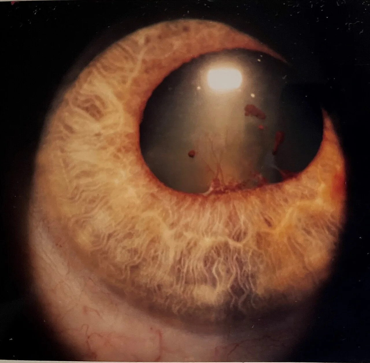
The iris root is its thinnest part and attaches to the ciliary body at the angle. It is the part that tears in blunt trauma, bleeding into the anterior chamber, and the part that bows forward against the cornea in angle closure.
What it does
The iris is an aperture. In bright light the sphincter constricts it, which cuts glare and increases depth of focus; in the dark the dilator opens it to let more light in. Both movements are reflexes, and both are a window on the nervous system. Light in either eye constricts both pupils, because the afferent signal from each retina is shared at the midbrain. Looking at a near object constricts the pupils too, together with convergence and accommodation.
The pupil's reactions are therefore an examination of the optic nerve, the midbrain, the third nerve and the sympathetic chain, all with a torch. A relative afferent pupillary defect, where the pupils dilate as the light swings from the good eye to the bad, is the single most useful sign of optic nerve disease.
How to examine it
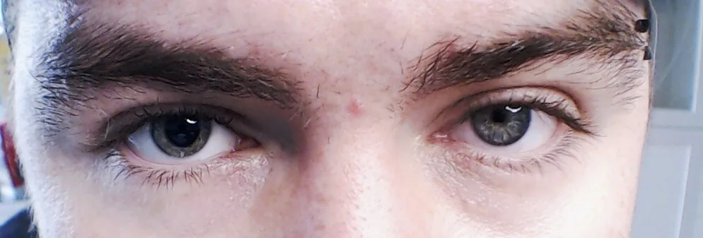
In diffuse light, look at the shape of the pupil, whether the two pupils are the same size in light and in dark, and the surface of the iris for crypts, new vessels and nodules. Then with the slit lamp narrow the beam and cross the pupil margin: a pupil stuck to the lens by posterior synechiae is irregular and does not dilate evenly. Retroillumination, the beam aimed through the pupil so the red reflex lights the iris from behind, shows holes in the iris as bright spots: atrophy from herpes zoster, a laser iridotomy, the transillumination of pigment dispersion.
A pupil that is peaked or torn after trauma points to the cornea or sclera: the iris has plugged a wound.
What goes wrong
Anterior uveitis, iritis, is inflammation of the iris and ciliary body: a deep aching photophobic red eye with a small pupil, cells in the anterior chamber, and a risk of synechiae. Rubeosis is new vessels growing on the iris when the retina is ischaemic, from diabetes or a vein occlusion, and it leads to a painful neovascular glaucoma when the vessels reach the angle. Iris melanoma is the rarest of the uveal melanomas and the most visible.
The pupil's own disorders are neurological. A dilated pupil with a droopy lid and an eye that cannot look in or up is a third nerve palsy, and with pain it is an aneurysm until imaged. A small pupil with a slight ptosis is Horner syndrome, and in a smoker it can be a lung apex tumour. A pupil that is large and reacts slowly to light but better to near is a tonic Adie pupil, benign and usually in a young woman.
Examine it
Posterior synechiae stick it to the lens in uveitis; its root forms the angle that closes in acute angle-closure glaucoma.
Ask OphthoSpace about the iris
AI responses are for education and may contain errors. OphthoSpace is not for primary clinical diagnosis.