Globe wall · 11 of 24
What is the sclera?
The sclera is the white of the eye: a tough coat of dense collagen that forms the outer wall of five sixths of the globe, continuous with the clear cornea in front at the limbus and with the sheath of the optic nerve behind. It holds the eye's shape under pressure, gives the six extraocular muscles something to pull on, and is pierced by every vessel and nerve that enters the eye.
- Thickness
- About 1 mm at the back, 0.3 mm just behind the muscle insertions, the thinnest point
- Made of
- Irregular collagen bundles, which is why it is white and the cornea is clear
- Openings
- The optic nerve behind, the ciliary arteries and nerves, the vortex veins at the equator
- Covered by
- Episclera, Tenon's capsule, and the conjunctiva in front
Three coats over the eye
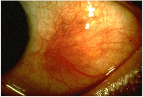
Three tissues lie over the sclera and each one can be red. The conjunctiva is the thin transparent mucous membrane on top, loosely attached, running from the lid margin into the fornix and onto the globe up to the limbus. Beneath it is Tenon's capsule and the episclera, a vascular connective tissue layer. Then the sclera itself, with its own deep vessels. Which layer's vessels are dilated decides whether a red eye is conjunctivitis, episcleritis or scleritis, and a drop of phenylephrine blanches the conjunctival and episcleral vessels but not the scleral ones.
At the limbus the sclera thins to a groove that holds Schlemm's canal, and the corneal stem cells live in the conjunctival border on top of it. Where the optic nerve leaves, the sclera becomes the lamina cribrosa, a perforated plate the nerve fibres pass through.
How to examine it
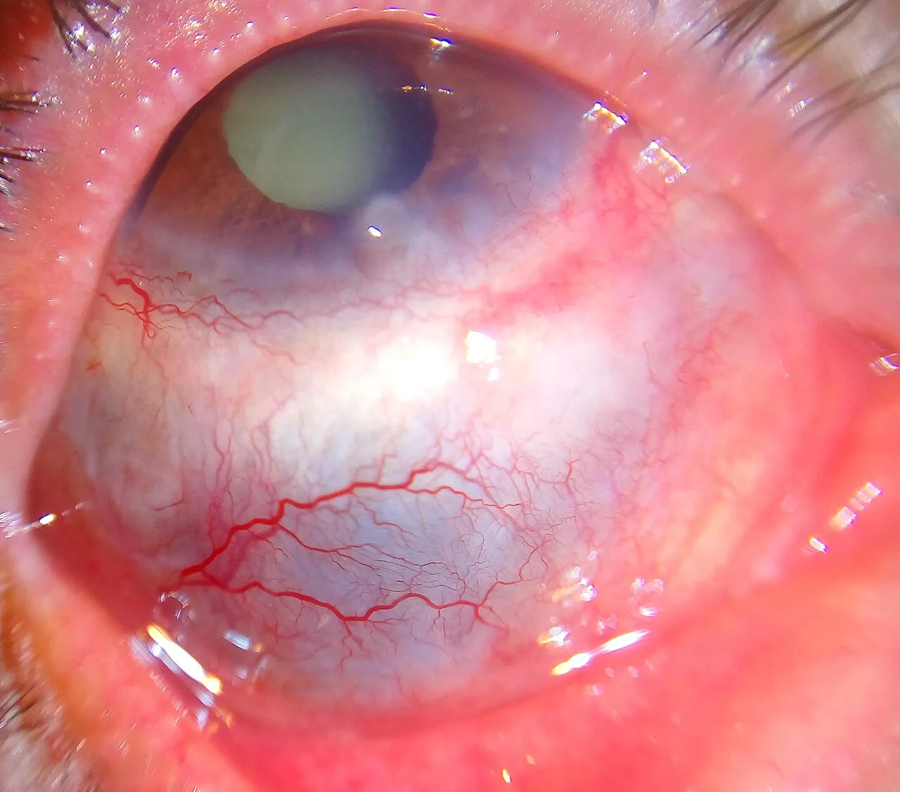
Look at the pattern of redness in daylight before any drops go in. Conjunctival injection is brightest in the fornices and fades towards the cornea, and the vessels move with the conjunctiva. Ciliary injection is a ring around the limbus and points to the cornea or the inside of the eye. Scleral injection is deep, bluish red and often in a sector, and the eye is tender to touch. Then look at the sclera in natural light for a blue or grey tint, which means it has thinned and the choroid shows through.
Episcleritis and scleritis
Episcleritis is a sectoral redness of the superficial episcleral vessels, mildly uncomfortable, self-limiting, and usually in a healthy young adult. Scleritis is different in kind: a deep, boring pain that wakes the patient at night and is worse on eye movement, a violaceous injection that does not blanch, and in half of patients a systemic disease, most often rheumatoid arthritis or a vasculitis. Necrotising scleritis thins the wall until the choroid shows through, and it can perforate. Scleritis needs systemic treatment and a search for its cause.
| Episcleritis | Scleritis | |
|---|---|---|
| Pain | Mild ache or grittiness | Deep, boring, wakes the patient, worse on movement |
| Colour | Bright red, superficial | Deep violaceous red |
| Phenylephrine | Blanches | Does not blanch |
| Tenderness | None or slight | Marked |
| Systemic disease | Uncommon | About half: rheumatoid, vasculitis, and others |
| Treatment | Lubricants, oral NSAID if needed | Systemic NSAID, steroid or immunosuppression |
Other things seen on the white of the eye
A subconjunctival haemorrhage is a flat bright red patch with a sharp edge, painless, from a small conjunctival vessel after coughing, straining or nothing at all; it clears in two weeks and needs only a blood pressure check. A blue sclera is thin sclera: osteogenesis imperfecta, or thinning after scleritis. A pterygium is a wing of conjunctiva growing onto the cornea from the nasal side in sun-exposed eyes. Scleral rupture after blunt trauma is easy to miss: a soft eye, a deep chamber, a dark bulge under the conjunctiva, and it needs a shield and a surgeon, not pressure.
Examine it
Scleritis is a deep, boring pain that wakes the patient; episcleritis is superficial and blanches with phenylephrine.
Ask OphthoSpace about the sclera
AI responses are for education and may contain errors. OphthoSpace is not for primary clinical diagnosis.