Lens · 4 of 24
What is the lens?
The lens is a transparent, flexible, biconvex disc about ten millimetres across and four millimetres thick, held behind the iris by the zonules. It adds the fine focus that the cornea's fixed power cannot: it rounds up to bring near objects onto the retina and flattens for distance. It keeps growing throughout life, laying new fibres over old, and that is why it stiffens in middle age and clouds in old age.
- Size
- About 10 mm across, 4 mm thick, growing slowly all life
- Power
- About 15 to 20 dioptres at rest, a third of the eye's total
- Accommodation
- Up to about 14 dioptres in a child, almost none after sixty
- Parts
- Capsule, cortex, nucleus; the anterior epithelium under the capsule makes new fibres for life
Capsule, cortex, nucleus
The lens is wrapped in a capsule, an elastic basement membrane that the zonules pull on. Beneath the front of the capsule is a single layer of epithelium, and at the equator those cells elongate into fibres and lay themselves down over the fibres before them. The oldest fibres are in the centre, the nucleus; the younger ones around it form the cortex. Because nothing is ever shed, the nucleus is made of cells from before birth, and it is the part that yellows and hardens with age.
The lens has no blood supply and no nerves. It lives on the aqueous, which is why it clouds when the aqueous chemistry changes: in diabetes, in hypocalcaemia, and after intraocular inflammation.
Accommodation and why it fails
At rest the zonules hold the lens under tension and flattened. To focus near, the ciliary muscle contracts, the ring it forms gets smaller, the zonules slacken, and the lens rounds up under the elasticity of its own capsule. That is accommodation, and it is coupled with convergence and pupil constriction in the near reflex.
As the nucleus hardens the lens can no longer round up, and reading arms grow longer. That is presbyopia, arriving in the mid forties in almost everyone, corrected simply by adding plus power for near. It is not a disease, and it is not the beginning of cataract.
How to examine it
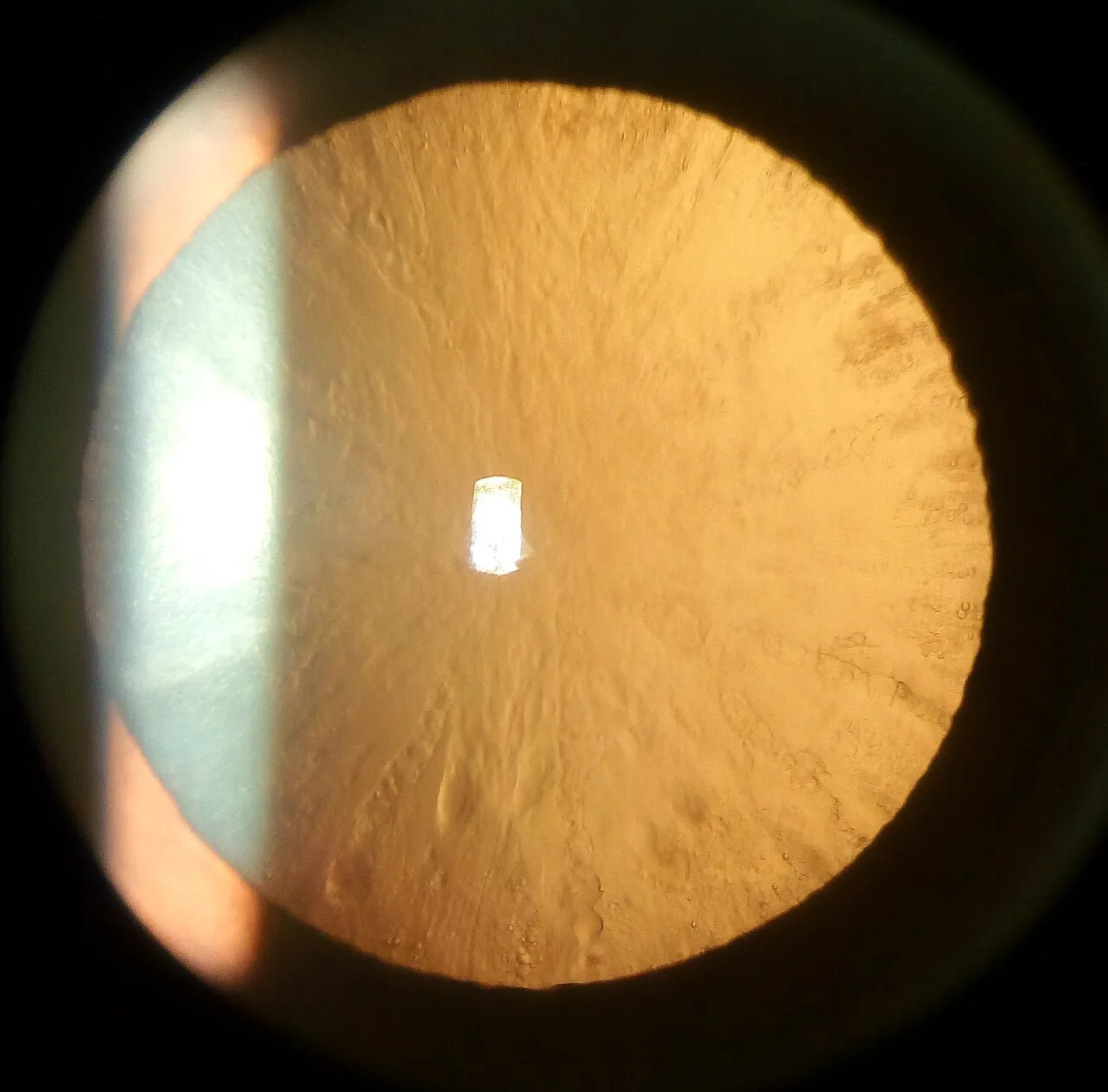
Two views show the lens. In the slit lamp's optical section, the lens is a series of layered bands behind the iris: the anterior capsule as a bright line, the cortex, the denser nucleus, the posterior capsule. Where each opacity sits in that section names the cataract. Then look at it with retroillumination: send the beam through the pupil, focus on the lens, and the orange glow from the fundus lights it from behind, so cortical spokes and posterior subcapsular plaques show as dark shapes against the red reflex.
With an ophthalmoscope held at arm's length, the red reflex does the same work in a moment: a bright even reflex means clear media, a dull or blotched one means something in the way, and a white reflex in a child is an emergency.
Cataract
A cataract is any opacity of the lens, and age is by far the commonest cause. Three patterns are worth telling apart, because they behave differently. Nuclear sclerosis is the yellowing and hardening of the centre: slow, symmetrical, and it shifts the eye towards short sight so that a patient may briefly read without glasses again. Cortical cataract is wedge-shaped spokes in the outer layers, which scatter light and cause glare from headlights. Posterior subcapsular cataract is a plaque on the back capsule, right on the visual axis, so it cuts near vision and vision in bright light early, and it is the pattern of steroids and diabetes.
Surgery removes the lens contents through a small incision by phacoemulsification and leaves the capsule to hold an artificial lens. It is done when the cataract limits what the patient wants to do, not at a threshold of acuity.
| Type | Where in the section | How it presents |
|---|---|---|
| Nuclear sclerosis | The central nucleus, yellow to brown | Slow blur, a myopic shift, dulled colours |
| Cortical | Spokes in the outer cortex | Glare and haloes, often little effect on acuity at first |
| Posterior subcapsular | A plaque on the back capsule | Early loss of reading and bright-light vision; steroids, diabetes, younger patients |
| Mature or white | The whole lens opaque | Hand movements vision, no red reflex; a swollen lens can close the angle |
When the lens moves
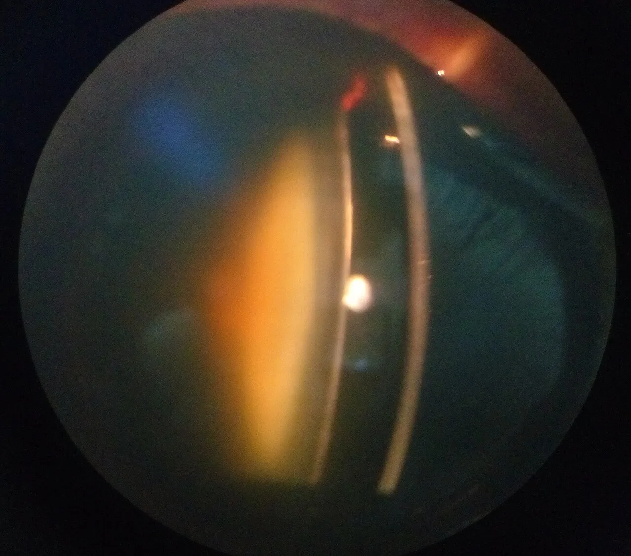
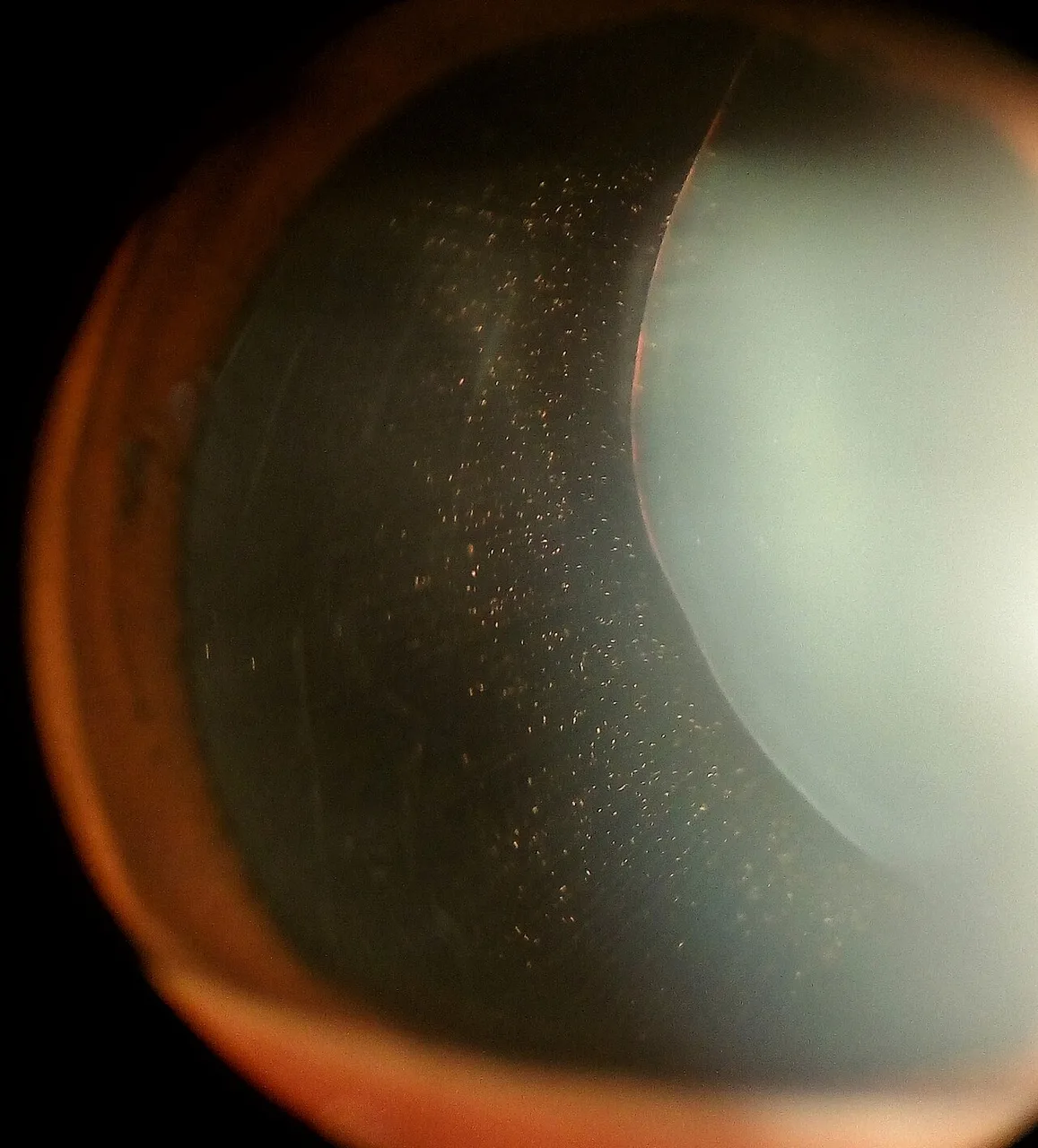
If the zonules are weak or broken the lens shifts, subluxation, or falls out of the pupil entirely. The causes are trauma, Marfan syndrome, where it goes up and out, homocystinuria, where it goes down and in, and pseudoexfoliation in older patients. The patient notices fluctuating vision and a doubling in one eye; at the slit lamp the edge of the lens crosses the dilated pupil and the iris trembles with eye movement.
Examine it
Cataract is the lens losing its clarity; refractive error is the lens and cornea missing the retina.
Ask OphthoSpace about the lens
AI responses are for education and may contain errors. OphthoSpace is not for primary clinical diagnosis.