Anterior segment · 5 of 24
What is the ciliary body?
The ciliary body is a ring of tissue running around the inside of the eye just behind the iris, between the iris root and the front edge of the retina. It is the middle part of the uvea, with the iris in front and the choroid behind. It does two jobs that nothing else can: it makes the aqueous humour that inflates the eye, and it focuses the lens.
- Width
- About 6 mm from the iris root back to the ora serrata
- Parts
- The pars plicata in front, with about seventy ciliary processes; the flat pars plana behind
- Muscle
- The ciliary muscle, parasympathetic through the third nerve via the ciliary ganglion
- Makes
- Aqueous humour at about 2 to 3 microlitres a minute
Processes and muscle
Seen from behind, the front part of the ciliary body is a ring of radial ridges, the ciliary processes, each one a fold of vascular stroma covered by a double layer of epithelium. That epithelium secretes the aqueous into the posterior chamber. The zonules attach in the valleys between the processes. Behind the processes is the pars plana, a smooth flat band that reaches back to the ora serrata where the retina begins. The pars plana is where the surgeon enters the eye, because there is no retina there to tear.
Inside the ciliary body is the ciliary muscle, a ring of smooth muscle in three orientations. When it contracts the ring narrows and moves forward, slackening the zonules so the lens can round up.
Aqueous production and pressure
The ciliary epithelium pumps aqueous into the posterior chamber by active secretion, a process that carbonic anhydrase drives and beta receptors stimulate. That is why acetazolamide and timolol lower pressure: they turn the tap down. The aqueous flows through the pupil, fills the anterior chamber and drains at the angle. Its production continues day and night, a little slower in sleep, so the eye's pressure is set by outflow.
Inflammation of the ciliary body reduces production, which is why an eye with severe uveitis can have a low pressure, and a long-standing very low pressure means the ciliary body has shut down or detached.
What goes wrong
Ciliary spasm is the aching pain of iritis and of a corneal abrasion: the muscle cramps, and a cycloplegic drop such as cyclopentolate relaxes it and relieves the pain. Intermediate uveitis is inflammation centred on the pars plana, with cells in the vitreous and snowbanks over the ora serrata, in a young patient with floaters. Ciliary body melanoma hides behind the iris and presents late, sometimes as a sentinel dilated vessel on the sclera over it.
In angle closure the ciliary body's position matters: a forward-rotated ciliary body or a plateau iris pushes the iris root into the angle even when the pupil block has been treated.
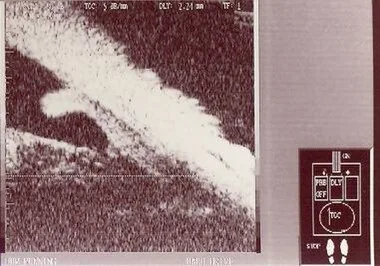
Examine it
It makes the aqueous that intraocular pressure depends on, and it is where intermediate uveitis and the pain of ciliary spasm arise.
Ask OphthoSpace about the ciliary body
AI responses are for education and may contain errors. OphthoSpace is not for primary clinical diagnosis.