Posterior segment · 10 of 24
What is the choroid?
The choroid is the layer of blood vessels and pigment between the retina and the sclera, the back part of the uvea. It has one of the highest blood flows per gram of any tissue in the body, and its innermost capillary bed, the choriocapillaris, feeds the photoreceptors through Bruch's membrane and the pigment epithelium. When it grows a vessel where it should not, the result is wet macular degeneration.
- Thickness
- About 0.2 to 0.3 mm, thickest under the macula, thinning with age and in myopia
- Layers
- Large vessels outside, medium vessels, then the choriocapillaris against Bruch's membrane
- Blood supply
- Short posterior ciliary arteries from the ophthalmic artery
- Feeds
- The outer retina: photoreceptors and the pigment epithelium
How it is built
From outside in, the choroid is the suprachoroidal space against the sclera, a layer of large vessels, a layer of medium vessels, and the choriocapillaris, a single flat sheet of wide fenestrated capillaries. Between the choriocapillaris and the pigment epithelium is Bruch's membrane, a five-layered sheet that thickens and stiffens with age. The choroid is full of melanocytes, which is why the fundus is orange rather than red and why in a lightly pigmented eye the large choroidal vessels show through as a tigroid pattern.
What it does
The choroid feeds the outer third of the retina, the photoreceptors and pigment epithelium, which the retinal vessels do not reach. It carries away the heat that focused light produces at the macula. Its pigment absorbs the light that passes through the retina so it is not scattered back. The fenestrated choriocapillaris leaks freely, and the pigment epithelium's tight junctions are the barrier that keeps that leak out of the retina: the outer blood-retinal barrier.
What goes wrong
Choroidal neovascularisation is a new vessel from the choriocapillaris breaking through Bruch's membrane and growing under the retina, where it leaks and bleeds. It is the mechanism of wet age-related macular degeneration, of the membranes that complicate high myopia and angioid streaks, and it is what anti-VEGF injections suppress. Central serous chorioretinopathy is a thick, leaky choroid pushing fluid through a pinpoint defect in the pigment epithelium to make a blister under the fovea.
Posterior uveitis and choroiditis are inflammation here: sarcoidosis, tuberculosis, toxoplasmosis and the white dot syndromes. Choroidal melanoma is the commonest primary eye cancer in adults, a pigmented dome under the retina, often with orange pigment on its surface. A choroidal detachment lifts the choroid and retina together as smooth brown mounds when the eye's pressure falls after surgery.
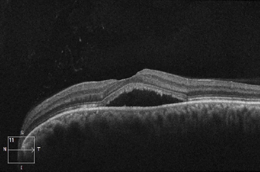
- 1Subretinal fluidAn optically empty (black) space between the neurosensory retina above and the RPE below: the retina has lifted off its pigment epithelium.
- 2Fovea, liftedThe foveal contour is preserved but the whole fovea sits on the dome of fluid, which is why the patient sees a grey smudge.
- 3Retinal pigment epitheliumThe bright continuous band. It is intact here; a break or a bump in it would point to a neovascular membrane instead.
- 4ChoroidThe mottled layer beneath the RPE. Thickened in central serous chorioretinopathy, which is part of why the fluid leaks.
Examine it
Choroidal neovascularisation is the wet form of macular degeneration; posterior uveitis and melanoma arise here.
Ask OphthoSpace about the choroid
AI responses are for education and may contain errors. OphthoSpace is not for primary clinical diagnosis.