Optic nerve · 12 of 24
What is the optic nerve?
The optic nerve carries every signal the eye produces: about 1.2 million ganglion cell axons running from the disc, through the orbit and the optic canal, to the chiasm, about fifty millimetres in all. It is not a peripheral nerve but a tract of the brain, myelinated by oligodendrocytes and wrapped in all three meninges, with cerebrospinal fluid in the subarachnoid space around it. That is why raised pressure in the head swells the disc, and why the diseases of the optic nerve are the diseases of white matter.
- Length
- About 50 mm: 1 mm in the eye, 25 mm in the orbit, 9 mm in the canal, 16 mm in the skull
- Fibres
- About 1.2 million, from the retinal ganglion cells
- Myelin
- Begins behind the lamina cribrosa, made by oligodendrocytes
- Blood
- Posterior ciliary arteries at the head; pial vessels along its length
From the disc to the chiasm
The nerve leaves the globe through the lamina cribrosa a little nasal to the back pole, runs an S-shaped course through the orbital fat inside the cone of the muscles, which gives the eye slack to move, and enters the skull through the optic canal alongside the ophthalmic artery. In the skull it runs back and medially to the chiasm, above the pituitary, where the fibres from the nasal half of each retina cross. So the temporal fields, which the nasal retinas see, cross at the chiasm, and a pituitary tumour pressing on it from below takes both temporal fields: a bitemporal hemianopia.
The fibres keep their retinal order along the nerve. The papillomacular bundle, from the macula, runs in the centre and is the first to suffer in toxic and nutritional neuropathies, which is why they take central vision and colour.
How to examine it
The optic nerve is examined by function first: acuity, colour vision with Ishihara plates, which the nerve loses before acuity, the pupils for a relative afferent defect, and the visual field by confrontation and on the perimeter. Then the disc, which is the only part of the nerve you can see. A relative afferent pupillary defect is the most objective sign of unilateral optic nerve disease and does not depend on the patient's cooperation. Imaging with MRI shows the nerve along its length; OCT measures the nerve fibre layer at the disc.
- Acuity, then colour, then pupils, then field, then the disc: the same order every time.
- Colour desaturation in one eye with a normal acuity is early optic neuropathy.
- An afferent pupillary defect with a normal disc means the lesion is behind the eye: retrobulbar neuritis or compression.
What goes wrong
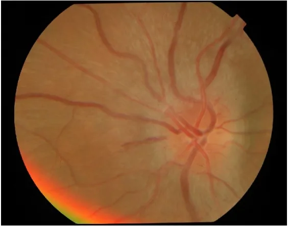
Optic neuritis is demyelination of the nerve, usually in a young woman: vision falls over days, it hurts to move the eye, colours wash out, and most recover in weeks. Half go on to multiple sclerosis, so the brain is imaged. Ischaemic optic neuropathy is an infarct of the nerve head: sudden painless loss, often on waking, with a swollen pale disc and an altitudinal field defect; in a patient over fifty with headache, jaw claudication or a tender temporal artery it is giant cell arteritis, and high dose steroids are given before the biopsy result to save the other eye. Compression by a tumour, an aneurysm or thyroid eye disease is slow and painless with a pale disc. Toxic and nutritional neuropathies from ethambutol, methanol, alcohol and B12 deficiency are bilateral and central.
Papilloedema is not a disease of the nerve but of the pressure around it: swollen discs in both eyes, vision preserved at first, from a mass, venous sinus thrombosis or idiopathic intracranial hypertension.
| Condition | Onset | Pain | Disc | Field |
|---|---|---|---|---|
| Optic neuritis | Days | On eye movement | Swollen in a third, normal in the rest | Central scotoma |
| Non-arteritic ischaemic | Sudden, on waking | None | Swollen, sectoral, small crowded fellow disc | Altitudinal |
| Arteritic ischaemic | Sudden | Headache, scalp, jaw | Swollen and pale | Altitudinal or total |
| Compression | Months | None | Pale, or swollen if in the orbit | Any; junctional at the chiasm |
| Papilloedema | Weeks | Headache | Swollen, both eyes | Enlarged blind spots, then constriction |
Beyond the chiasm
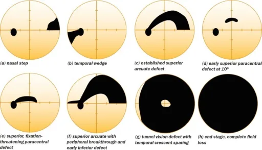
Behind the chiasm the fibres of each optic tract carry the same half of the visual field from both eyes, so every lesion from the tract to the occipital cortex makes a homonymous defect: the same side of the field in both eyes. The further back the lesion, the more alike the two eyes' defects, congruous, and the more likely the macula is spared. A field defect that respects the vertical midline is behind the eye; one that respects the horizontal is in the eye.
Examine it
Optic neuritis, ischaemic optic neuropathy and papilloedema each mark it in their own way; a relative afferent pupillary defect is its signature.
Ask OphthoSpace about the optic nerve
AI responses are for education and may contain errors. OphthoSpace is not for primary clinical diagnosis.