Interpret / Fundus
Disc. Vessels. Macula. Periphery.
Every fundus photograph is read in the same order so nothing is missed. Learn the order, describe the disc feature by feature, put normal beside abnormal, and then read the atlas, where the diagnosis stays hidden until you commit.
1. Optic disc
Assess colour, margins, and the cup-to-disc ratio. Look for pallor, swelling, haemorrhage, and new vessels at the disc (NVD).
2. Vessels
Follow the arcades out from the disc. Note arteriovenous ratio, nicking, tortuosity, emboli, sheathing, and neovascularization elsewhere (NVE).
3. Macula
Inspect the fovea and surrounding retina for drusen, exudates, haemorrhage, oedema, pigment change, and a foveal light reflex.
4. Periphery
Sweep the four quadrants for tears, detachment, lattice, pigmentary change, and peripheral haemorrhage or exudate.
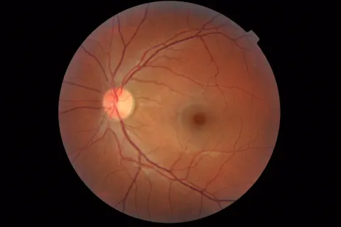
Describe the disc
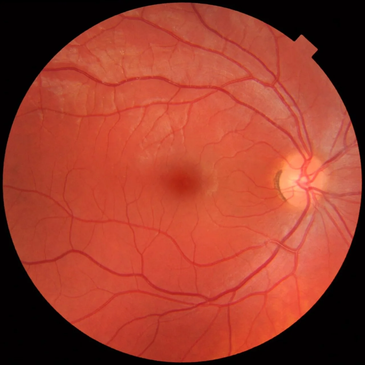
Mikael Häggström, CC0, source.
Describe the disc
Margins
Colour
Cup to disc ratio
Swelling
Haemorrhage
Vessels at the disc
Choose each feature to build the description.
Normal beside abnormal
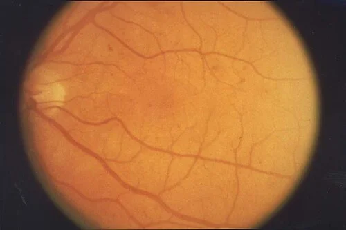
The words
- Microaneurysm
- A tiny red dot, the earliest sign of diabetic retinopathy: a bulge in a capillary wall.
- Dot and blot haemorrhage
- Round haemorrhages in the deep retina, where the cells are packed vertically.
- Flame haemorrhage
- Feathery, in the nerve fibre layer, following the fibres: hypertension, vein occlusion, papilloedema.
- Hard exudate
- Yellow, waxy, sharp-edged lipid left behind by leaking vessels, often ringed around the leak.
- Cotton wool spot
- White, fluffy, soft-edged: an infarct of the nerve fibre layer.
- Drusen
- Yellow deposits under the RPE at the macula; the substrate of age-related macular degeneration.
- Neovascularisation
- Fine, lacy new vessels on the disc (NVD) or elsewhere (NVE): the retina is ischaemic.
- Cup to disc ratio
- The pale central cup as a fraction of the disc diameter; above 0.6, or asymmetric between eyes, think glaucoma.
- Arteriovenous nicking
- A vein pinched where a stiff arteriole crosses it: hypertensive change.
- Cherry red spot
- A red fovea against a pale, oedematous retina: central retinal artery occlusion.
The fundus atlas
All 39 in the atlas
A routine fundus photograph at diabetic screening
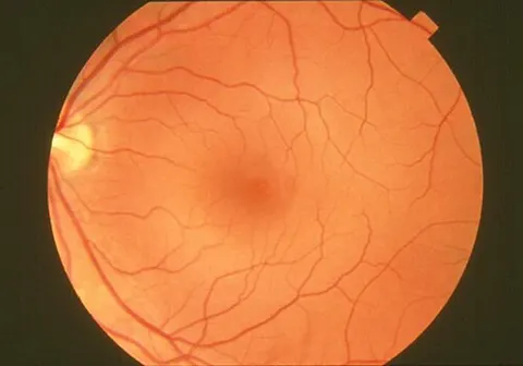
Fundus photograph before a hypertension clinic review
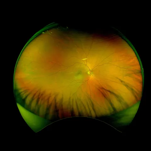
Ultra-widefield image at a myopia review
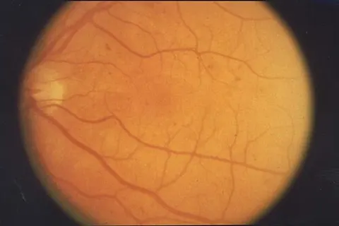
Screening photograph in a patient with type 2 diabetes
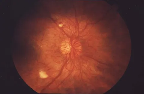
Floaters in a patient with long-standing type 1 diabetes
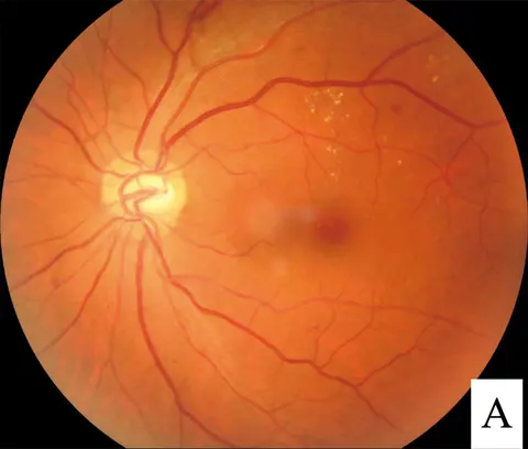
Gradual central blur in a patient with diabetes
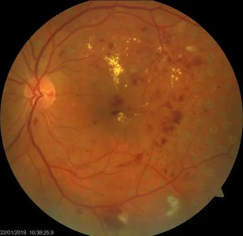
Distortion after previous retinal laser
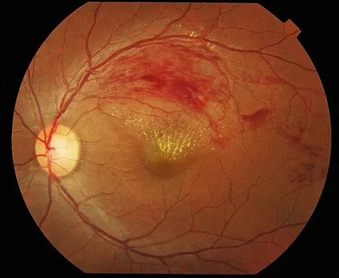
Painless blur in one eye in a hypertensive adult
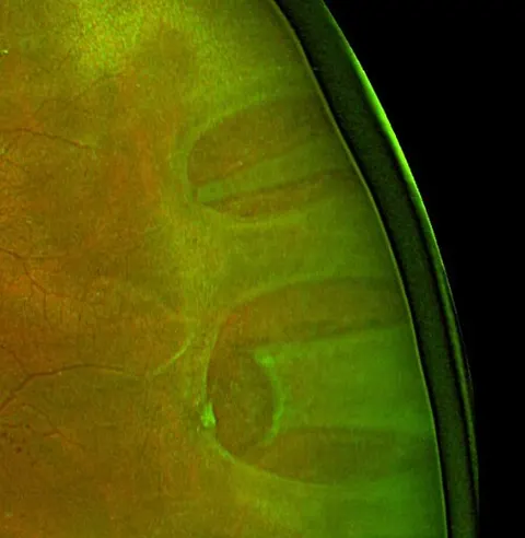
Flashes, floaters and a shadow in a myope
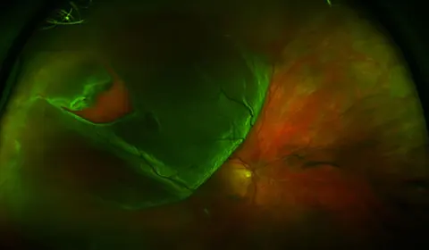
A curtain over the vision since yesterday
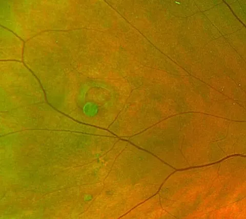
An incidental finding at a dilated examination
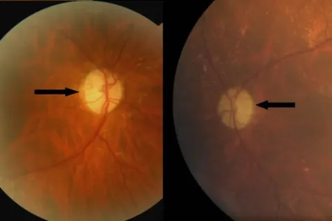
Poor colour vision in a young adult with a family history
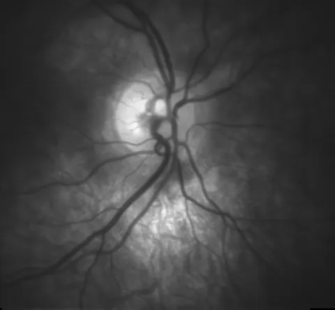
Red-free disc photograph of a glaucoma suspect
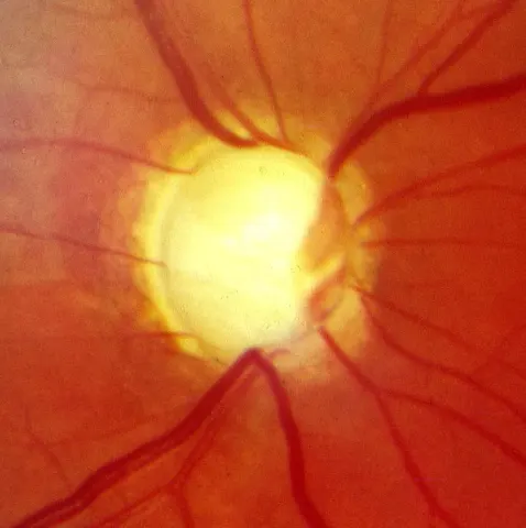
A disc found at a routine sight test, in a patient with no symptoms
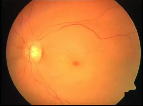
The same emergency in another eye
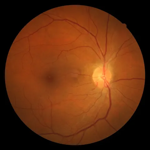
The fundus of a 58-year-old with a blood pressure of 175/105
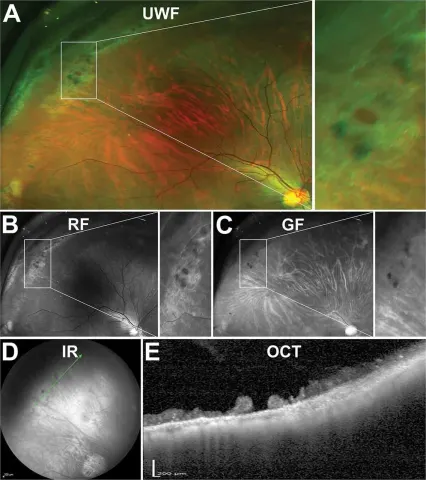
An atrophic hole within lattice, on every modality
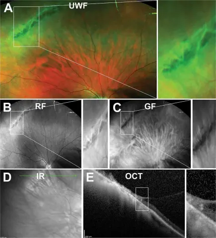
Criss-cross white lines in the far periphery of a 60-year-old
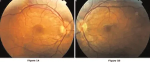
Swollen discs in both eyes of a young man with mouth ulcers
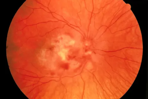
White necrosis and haemorrhage beside the disc in a transplant patient
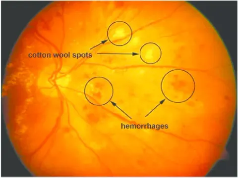
Cotton wool spots and haemorrhages, labelled
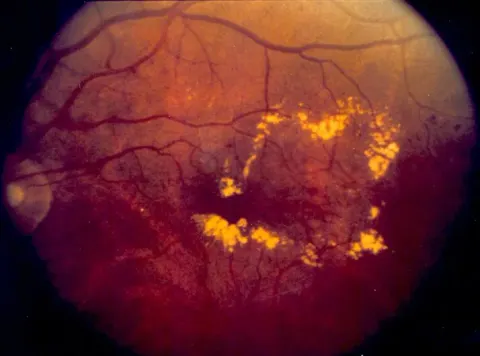
Blurred reading in a diabetic, with rings of yellow around the macula
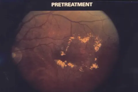
Diabetic macular oedema before treatment
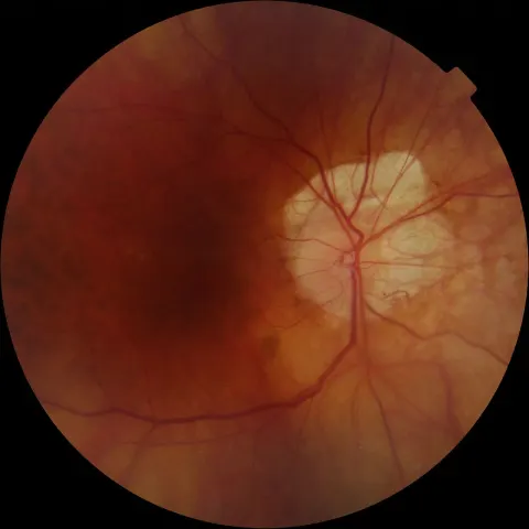
A pale halo around the disc of a very short-sighted right eye
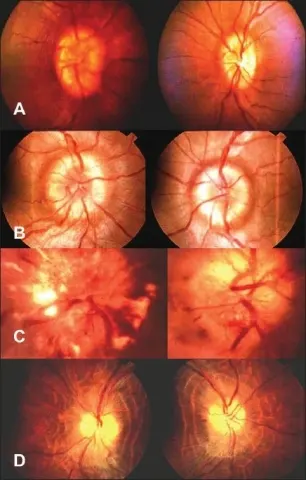
Papilloedema from early to atrophic, in four panels
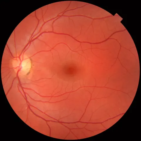
The fellow eye at diabetic screening
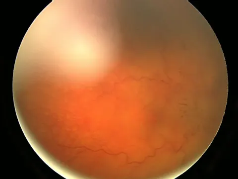
Headache worse on waking, and a photograph of the right disc
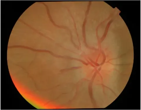
Pain on moving the right eye, then the vision went
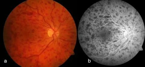
Sudden painless blur in one eye of a hypertensive 70-year-old
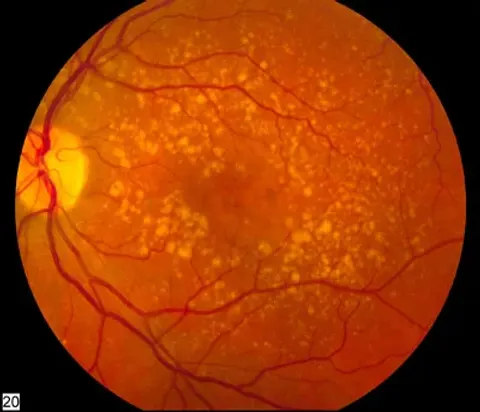
Reading is slower than it was, at 74
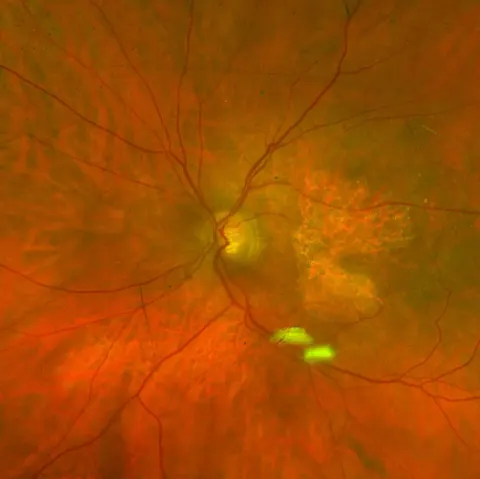
A blank patch in the middle of the page
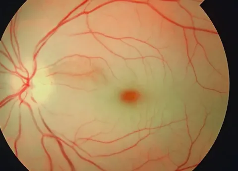
Vision gone in seconds, painless, an hour ago
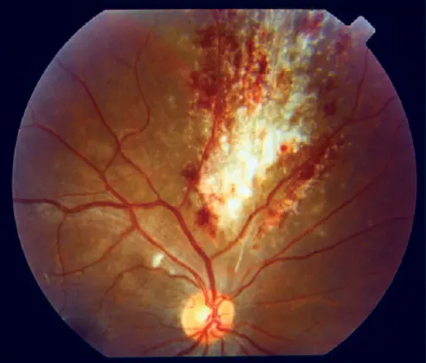
Floaters and blur in a patient with a very low CD4 count
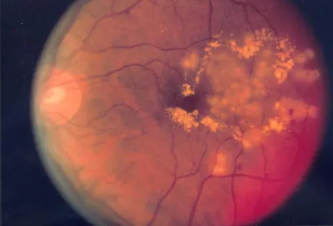
Diabetic maculopathy, after the laser
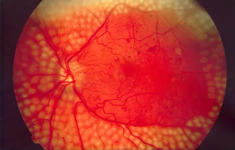
The retina after panretinal photocoagulation
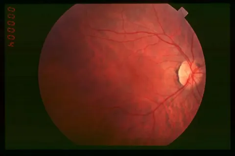
Trouble seeing in the dark since the teens: the early fundus
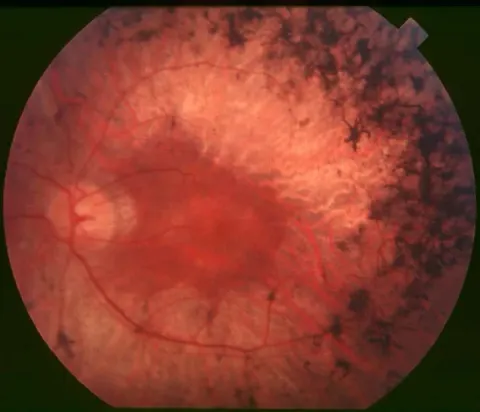
Bumping into things at the sides: the established fundus
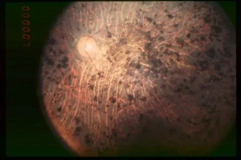
Registered blind at 60: the end-stage fundus
A short-sighted 23-year-old with a normal examination
132 images in the whole atlas, every one checked on its source page.