Presentations / Gradual vision loss
Gradual vision loss
Weeks to years: the media, the macula, the nerve, or the glasses.
Slow loss is common and mostly treatable. It comes from the optics (refractive error), the media (cataract), the macula (degeneration, diabetic maculopathy) or the nerve (glaucoma, a compressive lesion). The pinhole and the pattern of loss sort them.
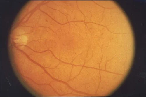
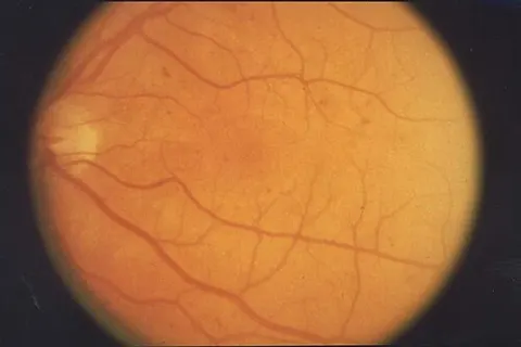
Screening photograph in a patient with type 2 diabetesOpen in the atlas
The four places
Improves with a pinhole: refractive. A dulled red reflex and glare: cataract. Distortion and central loss with a normal periphery: the macula. Peripheral loss the patient did not notice, with a cupped disc: glaucoma. Colour loss, an afferent defect and a field respecting the vertical: the nerve or the chiasm, and an image of the brain.
| Cause | The clue | The finding | What happens next |
|---|---|---|---|
| Refractive error and presbyopia | Better through a pinhole; reading at arm's length | Normal eye | Glasses |
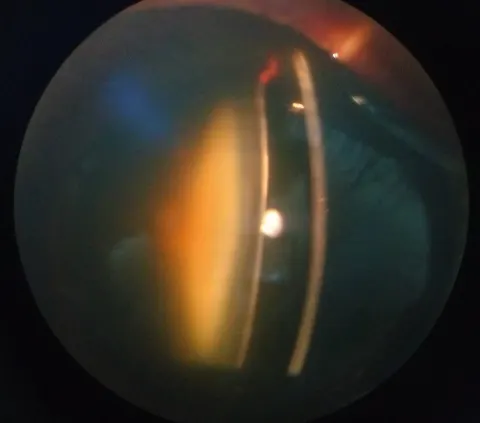
| Cataract | Glare, dulled colours, a myopic shift | Dim red reflex, lens opacity | Surgery when it limits life |
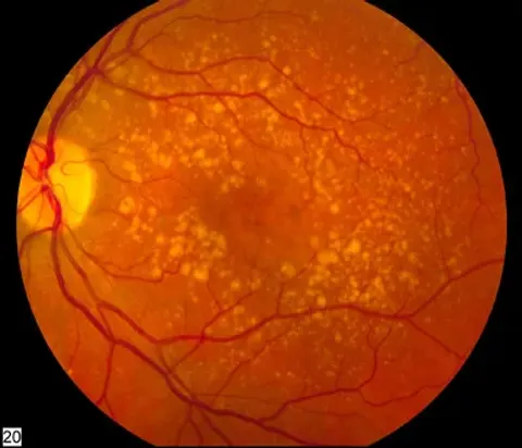
| Dry macular degeneration | Slow central blur, older patient | Drusen, atrophy | Vitamins, monitoring, an Amsler grid for the wet conversion |
| Diabetic maculopathy | Diabetes, gradual central blur | Exudate and thickening at the macula | Anti-VEGF or laser; OCT to follow |
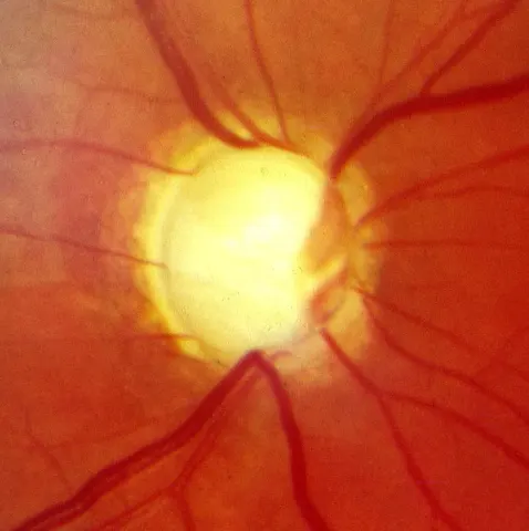
| Open-angle glaucoma | Nothing, until late | Cupped disc, raised pressure, arcuate field loss | Pressure-lowering drops for life |
| Compressive optic neuropathy | Colour loss, a field respecting the vertical, sometimes proptosis | Pale disc, RAPD | MRI of the orbit and chiasm |
The one that is a tumour
A pituitary adenoma presses on the chiasm from below and takes the temporal fields of both eyes, slowly, so the patient bumps into doorframes and blames clumsiness. Confrontation fields with a red target across the vertical midline find it in a minute. Image the sella.
Go and look
Where this topic leads