Posterior segment · 6 of 24
What is the vitreous?
The vitreous is the clear gel that fills the back four fifths of the eye, from the lens to the retina. It is ninety-nine per cent water held in a fine scaffold of collagen and hyaluronan, so it is transparent and nearly weightless, and it is attached to the retina most firmly at its base near the ora serrata, around the optic disc, and at the macula. Those attachments are where the trouble starts when it ages.
- Volume
- About 4 ml, most of the eye
- Made of
- Water, collagen fibrils and hyaluronan, with almost no cells
- Attachments
- The vitreous base at the ora serrata (strongest), the disc margin, the macula, and along vessels
- Renewal
- None: the vitreous is formed once and is never replaced
What it does
The vitreous holds the shape of the eye during growth, keeps the retina pressed against the choroid, and lets light through unscattered. It is also a metabolic buffer between the lens and the retina. Once the eye is grown it does not need to be a gel, which is why the eye copes with its removal at vitrectomy as long as it is refilled with fluid or gas.
How it ages, and the flashes and floaters that follow
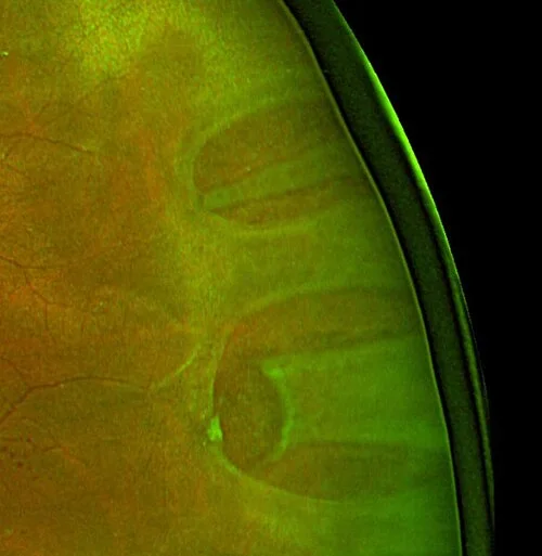
From middle age the gel liquefies in pockets and the collagen clumps into strands. Those strands cast shadows on the retina: floaters, seen best against a bright sky. Eventually the vitreous shrinks away from the back of the eye and separates from the retina, a posterior vitreous detachment. As it peels, it tugs on the retina and the tug is seen as a flash of light in the temporal field. The ring of attachment around the disc comes away as a Weiss ring, a single large floater.
In most eyes this is harmless. But where the vitreous is stuck to a weak spot in the retina the pull can tear it, and fluid then passes through the tear and lifts the retina off. That is why a new shower of floaters, flashes, or a shadow in the field is examined the same day: a tear treated with laser is a small matter, a detachment is an operation.
- Sudden floaters or flashes: dilated examination within twenty four hours to look for a tear.
- A curtain or shadow across the field means the retina is detaching; same day referral.
- Pigment in the anterior vitreous (tobacco dust) after a vitreous detachment means a tear until proved otherwise.
What else fills it
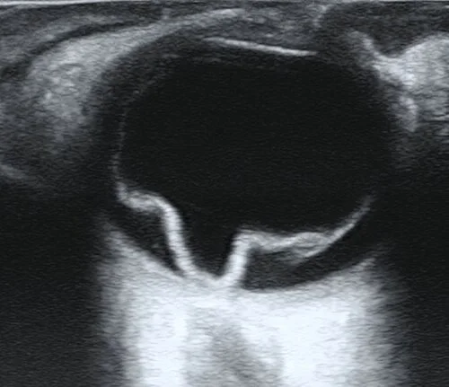
Blood in the vitreous, a vitreous haemorrhage, comes from new vessels in proliferative diabetic retinopathy, from a torn retinal vessel, or from a vein occlusion: sudden painless loss of vision with a dull red reflex. Inflammatory cells in the vitreous are vitritis, seen in intermediate and posterior uveitis and, after eye surgery or in a septic patient, in endophthalmitis, which is an emergency. When the fundus cannot be seen through the vitreous, B-scan ultrasound shows what lies behind it.
At the macula the vitreous can pull without letting go, vitreomacular traction, and the tug can open a full thickness macular hole.
Examine it
As it liquefies with age it can pull away from the retina: flashes, floaters, and sometimes a retinal tear.
Ask OphthoSpace about the vitreous
AI responses are for education and may contain errors. OphthoSpace is not for primary clinical diagnosis.