Posterior segment · 7 of 24
What is the retina?
The retina is the light-sensitive lining of the back of the eye, a sheet of nervous tissue about a quarter of a millimetre thick that turns light into signals and does the first stages of seeing before anything reaches the brain. It is part of the central nervous system, grown out from the forebrain, which is why it does not regenerate and why its diseases are so often permanent.
- Thickness
- About 0.2 to 0.3 mm, thinnest at the fovea
- Photoreceptors
- About 120 million rods for dim light and 6 million cones for colour and detail
- Blood supply
- Inner half from the central retinal artery; outer half, the photoreceptors, from the choroid
- Edge
- The ora serrata, where the retina ends against the ciliary body
Ten layers, and why the order matters
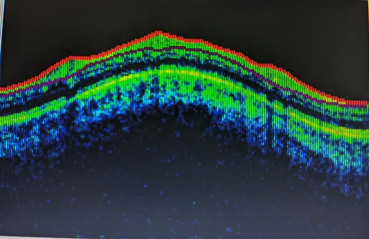
Light enters the retina from the inside of the eye and must pass through all of its layers before it reaches the photoreceptors, which lie at the back against the pigment epithelium. From inside out: the internal limiting membrane, the nerve fibre layer carrying the ganglion cell axons to the disc, the ganglion cell layer, the inner plexiform layer, the inner nuclear layer of bipolar and horizontal cells, the outer plexiform layer, the outer nuclear layer of photoreceptor cell bodies, the photoreceptor inner and outer segments, and the retinal pigment epithelium. Below the RPE is Bruch's membrane and then the choroid.
The order explains the diseases. Blood from the retinal circulation collects in the inner layers: flame haemorrhages track along nerve fibres, dot and blot haemorrhages sit in the nuclear layers. Exudate settles in the outer plexiform layer, in a star around the fovea. The photoreceptors depend on the choroid, so when the retina detaches from the RPE they starve, and a macula off for more than a few days does not fully recover.
The map of the fundus
Looking in, the optic disc is the pale round nerve head on the nasal side, and the macula is the darker area two disc diameters temporal to it, with the fovea at its centre. The vascular arcades curve above and below the macula from the disc. Everything beyond the arcades is the periphery, out to the ora serrata, which the direct ophthalmoscope never reaches and the widefield camera does.
Learn to read a fundus in one order every time: the disc, then the vessels out along each arcade, then the macula, then the four quadrants of periphery. A photograph is read the same way.
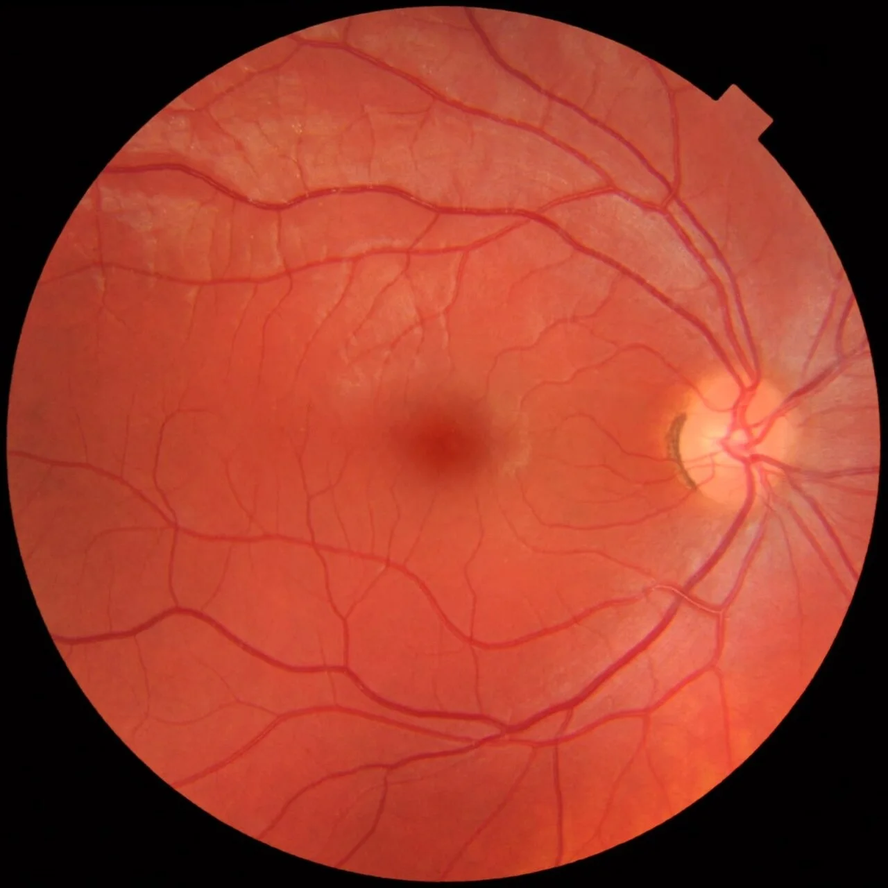
- 1Optic discPink rim, sharp margin, vessels leaving from the centre. About 1.5 mm across; a right eye, so the disc is on the right.
- 2CupThe pale central depression. Here about a third of the disc: a cup to disc ratio of 0.3 is normal.
- 3Central vesselsThe central retinal artery (narrower, brighter) and vein (wider, darker) emerge from the cup.
- 4Superotemporal arcadeThe main artery and vein arch above the macula. Follow each arcade out to the periphery.
- 5Inferotemporal arcadeThe matching arcade below. The macula lies between the two.
- 6MaculaThe darker central area between the arcades, two disc diameters temporal to the disc. No large vessels cross it.
- 7FoveaThe centre of the macula: darkest, avascular, with a small light reflex in a young eye.
How to examine it
The direct ophthalmoscope gives a magnified, upright view of a small field, best through a dilated pupil in a dark room. Find the red reflex from arm's length, come in close, find a vessel and follow it back to the disc. The slit lamp with a handheld lens gives a wide, inverted, stereoscopic view, and it is how the macula is properly seen. Fundus photography records the posterior pole; widefield imaging reaches the periphery; OCT sections the retina in cross section at the resolution of its layers.
Fluorescein angiography shows the vessels themselves: where they leak, where they have closed, and where new ones have grown.
What goes wrong
Retinal disease is mostly vascular, degenerative or mechanical. Diabetic retinopathy is the leading cause of blindness in working age: microaneurysms and haemorrhages first, then ischaemia, then new vessels that bleed. Hypertension narrows the arterioles and nicks the veins where they cross. A vein occlusion floods a sector or the whole retina with haemorrhage; an artery occlusion turns it pale with a cherry red spot at the fovea and is an eye stroke. Retinal detachment lifts the retina off its blood supply after a tear. Retinitis pigmentosa is the commonest inherited retinal degeneration: night blindness first, then tunnel vision, with bone spicule pigment in the periphery.
| Pattern | Look for | Think of |
|---|---|---|
| Dot and blot haemorrhages, microaneurysms | Small red dots near the macula | Diabetic retinopathy |
| Flame haemorrhages, cotton wool spots | Along the nerve fibres near the disc | Hypertension, vein occlusion, papilloedema |
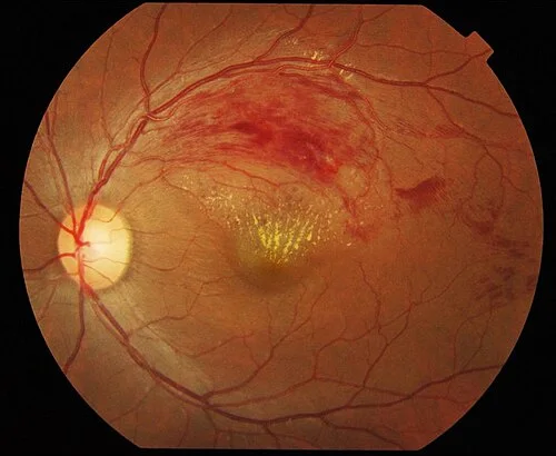
| Haemorrhage in one sector along a vein | A wedge of blood from an arteriovenous crossing | Branch retinal vein occlusion |
| Pale retina, cherry red fovea | A white posterior pole with a red centre | Central retinal artery occlusion: an emergency |
| Grey, wrinkled, elevated retina | Loss of the choroidal pattern; a tear at the edge | Retinal detachment |
| Bone spicule pigment in the mid periphery | Pale disc, thin arterioles | Retinitis pigmentosa |
Examine it
Diabetic and hypertensive retinopathy, vein and artery occlusions, and detachment are all read on its surface.
Ask OphthoSpace about the retina
AI responses are for education and may contain errors. OphthoSpace is not for primary clinical diagnosis.