Optic nerve · 9 of 24
What is the optic disc?
The optic disc is the head of the optic nerve, the place where a million nerve fibres from the whole retina gather and turn to leave the eye through a sieve in the sclera, the lamina cribrosa. It is about one and a half millimetres across, a little taller than it is wide, and it sits nasal to the macula. It has no photoreceptors, so it is the blind spot. Its appearance, the colour of its rim, the size of its cup, the sharpness of its edge, is the examination of the optic nerve.
- Size
- About 1.5 mm across, 1.75 mm tall; the unit of fundus distance is one disc diameter
- Position
- Nasal to the macula, by about two disc diameters
- Fibres
- About 1.2 million ganglion cell axons
- Vessels
- The central retinal artery and vein enter and leave at its centre
Rim, cup and margin
The disc is a pink ring of nerve fibres, the neuroretinal rim, around a pale central depression, the cup, from which the vessels emerge. The cup is where there are no fibres, just the lamina; its size in health depends on the size of the disc, so a big disc has a big cup. The ratio of cup to disc diameter is written down at every examination, and in a normal eye it is usually below half, with the two eyes within a tenth of each other.
The rim in a healthy disc obeys the ISNT rule: thickest inferiorly, then superiorly, then nasally, thinnest temporally. A rim that breaks the rule, notched inferiorly or superiorly, is losing fibres.
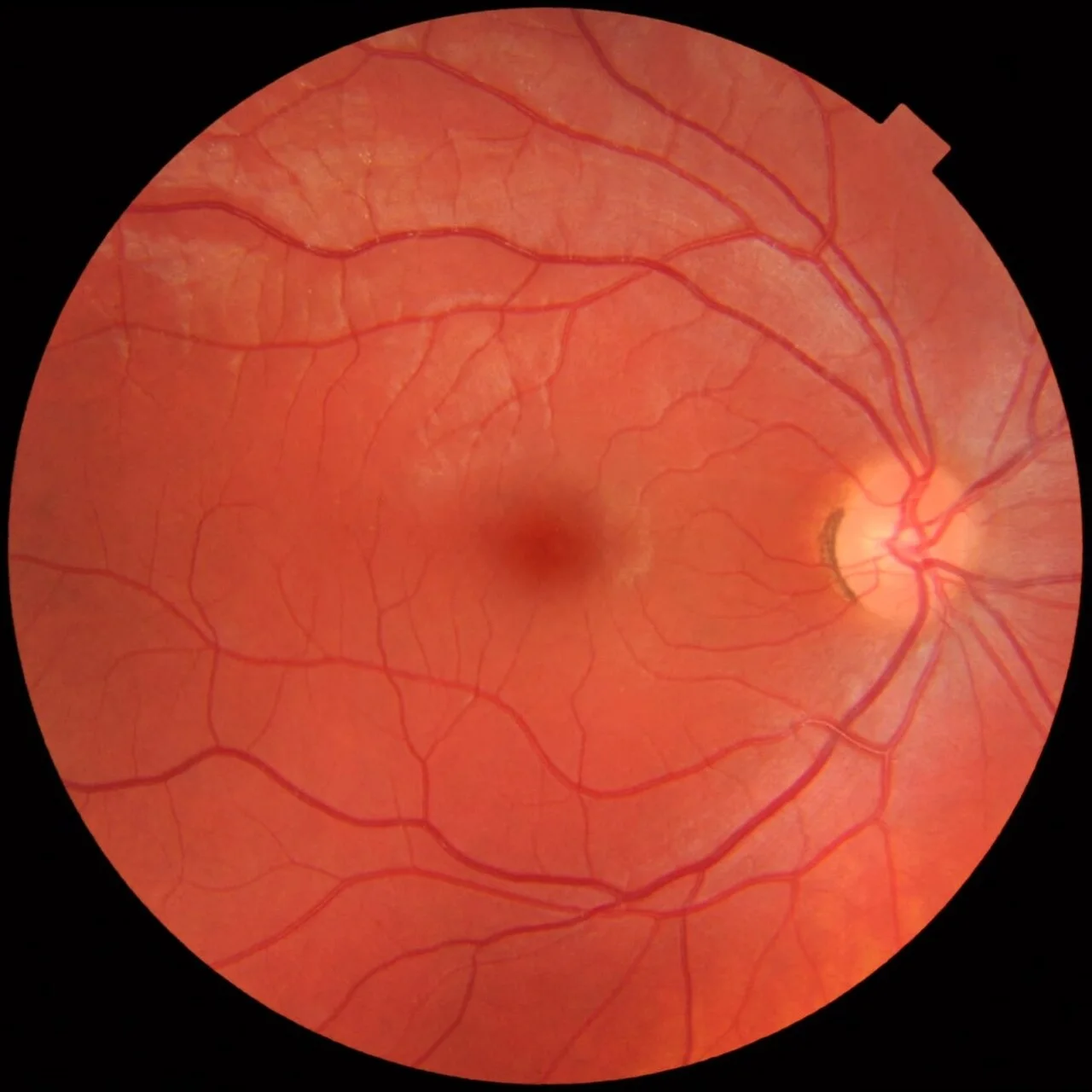
- 1Optic discPink rim, sharp margin, vessels leaving from the centre. About 1.5 mm across; a right eye, so the disc is on the right.
- 2CupThe pale central depression. Here about a third of the disc: a cup to disc ratio of 0.3 is normal.
- 3Central vesselsThe central retinal artery (narrower, brighter) and vein (wider, darker) emerge from the cup.
- 4Superotemporal arcadeThe main artery and vein arch above the macula. Follow each arcade out to the periphery.
- 5Inferotemporal arcadeThe matching arcade below. The macula lies between the two.
- 6MaculaThe darker central area between the arcades, two disc diameters temporal to the disc. No large vessels cross it.
- 7FoveaThe centre of the macula: darkest, avascular, with a small light reflex in a young eye.
How to examine it
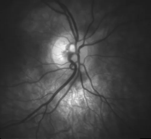
With the direct ophthalmoscope, find a vessel and follow it against its branching until it leads you to the disc. Judge the four things in order: colour of the rim, cup to disc ratio, the margin, and the vessels at the edge. A pale rim is atrophy; a large or asymmetric cup is glaucoma; a blurred margin with an elevated disc is swelling; new vessels on the disc are proliferative retinopathy. Red-free (green) light makes the nerve fibre layer visible as fine striations that fade in wedge defects.
OCT of the nerve head measures the thickness of the nerve fibre layer around the disc, and it finds glaucomatous loss before the field changes.
The cupped disc
Glaucoma is a slow death of ganglion cells, and it hollows the disc: the cup enlarges, the rim thins, vessels bend sharply over the edge of the cup and disappear into it, and a splinter haemorrhage may sit on the rim. Because the fibres from the arcuate bundles are lost first, the field loss is an arcuate scotoma with a nasal step, and the patient notices nothing until late. Pressure is the treatable risk factor, not the definition: a third of glaucoma is at normal pressure.
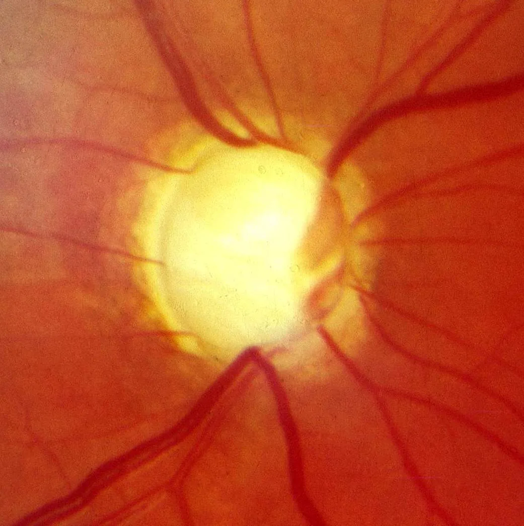
- 1The cupPale and wide: most of the disc is cup. A cup to disc ratio near 0.8 in a disc this size is glaucoma until proved otherwise.
- 2Thinned rimWhat is left of the neuroretinal rim: a narrow band of pink at the edge. In health the rim is thickest inferiorly, then superiorly, nasally, temporally (ISNT).
- 3Vessels bend at the edgeThe vessels dip over the lip of the cup and reappear on its floor, the bayoneting of a deep cup.
- 4Nasal shift of vesselsThe central vessels are pushed to the nasal edge as the cup enlarges.
The swollen disc
A swollen disc has blurred margins, is elevated, and buries the vessels at its edge under haemorrhages and cotton wool spots. Bilateral swelling from raised intracranial pressure is papilloedema, and the vision is normal at first, with headache and transient obscurations; it needs urgent imaging. Unilateral swelling with pain on eye movement and a loss of colour vision is optic neuritis. Sudden painless swelling in an older patient is ischaemic optic neuropathy, and if the temporal artery is tender it is giant cell arteritis, which blinds the other eye within days unless steroids start now.
A pale, flat, sharply-edged disc is atrophy: the end state of any of these, of long-standing compression, and of inherited optic neuropathies.
| Disc | Vision | Eyes | Think of |
|---|---|---|---|
| Swollen, both | Normal, then obscurations | Both | Papilloedema: image the head today |
| Swollen, one | Reduced, colours washed out, pain on movement | One | Optic neuritis |
| Swollen pale, one | Sudden altitudinal loss, no pain | One | Ischaemic optic neuropathy; check ESR and CRP for arteritis |
| Cupped | Normal until late | Both, asymmetric | Glaucoma |
| Pale, flat | Reduced, an afferent pupil defect | One or both | Optic atrophy |
Examine it
Cupping means glaucoma; swelling means papilloedema or optic neuritis; pallor means atrophy.
Ask OphthoSpace about the optic disc
AI responses are for education and may contain errors. OphthoSpace is not for primary clinical diagnosis.