Posterior segment · 8 of 24
What is the macula?
The macula is the centre of the retina, a disc about five and a half millimetres across, temporal to the optic disc and between the vascular arcades. At its centre is the fovea, a pit half a millimetre wide packed with cones and with no rods and no vessels at all. Everything you read, every face you recognise, is seen with the fovea; the rest of the retina tells you where to look.
- Size
- About 5.5 mm across; the fovea about 1.5 mm, the foveola 0.35 mm
- Position
- Two disc diameters temporal to the disc and slightly below its centre
- Cells
- The highest density of cones in the retina, and no rods at the very centre
- Blood
- No retinal vessels over the fovea; it lives on the choroid beneath
Why it looks dark, and why there is a pit
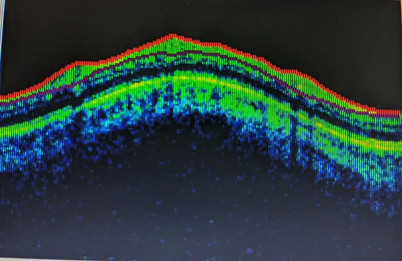
The macula is darker than the surrounding retina because of the yellow pigments lutein and zeaxanthin in its inner layers, which is where the name comes from, macula lutea, the yellow spot, and because the pigment epithelium beneath it is denser. At the fovea the inner retinal layers are swept aside so that light reaches the cones with nothing in the way: on OCT that is the foveal pit, and in a young eye its walls reflect the ophthalmoscope light as a bright foveal reflex.
The cones here are packed so tightly and wired so directly, one cone to one ganglion cell, that the fovea gives the eye its full acuity. Move one degree away and acuity halves.
How to examine it
Ask the patient to look straight at the light and the fovea is in the centre of your view. With the direct ophthalmoscope it is the last thing to look at, because the light constricts the pupil. The slit lamp with a lens shows it in stereo, which is how oedema and a hole are seen as elevation and depth. An Amsler grid in the patient's hand finds distortion before any of this: straight lines that bend mean the fovea has moved.
OCT is the macula's own examination. It shows the pit, each layer, and every compartment fluid can sit in, and it has made the difference between the kinds of macular disease routine to see.
What goes wrong
Macular disease takes central vision and leaves the periphery: the patient cannot read or see faces but walks in without help. Age-related macular degeneration is the commonest cause of blindness in the developed world. Its dry form is drusen, yellow deposits under the pigment epithelium, progressing over years to geographic atrophy; its wet form is a new vessel growing up from the choroid through Bruch's membrane, which leaks and bleeds and destroys vision in weeks unless injected. Diabetic macular oedema is leaking capillaries thickening the retina with exudate in rings. Central serous chorioretinopathy is a blister of fluid under the fovea in a young stressed man. A macular hole is a full thickness defect at the fovea pulled open by the vitreous. An epiretinal membrane is a sheet of scar on the surface that wrinkles the retina and distorts lines.
| Condition | On the fundus | On OCT |
|---|---|---|
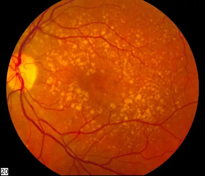
| Dry AMD | Drusen, pigment change, patches of atrophy | Bumps under the RPE; thinning where atrophy has occurred |
| Wet AMD | Grey membrane, haemorrhage, exudate at the fovea | Subretinal or intraretinal fluid with a break or bump in the RPE |
| Diabetic macular oedema | Rings of hard exudate, thickened retina | Cystoid spaces in the inner layers |
| Central serous | A shallow round blister at the macula | Subretinal fluid under an intact RPE, thick choroid |
| Macular hole | A round red defect at the fovea | A full thickness gap with cystic edges |
| Epiretinal membrane | A glistening surface with wrinkled vessels | A bright line on the surface with the retina puckered beneath |
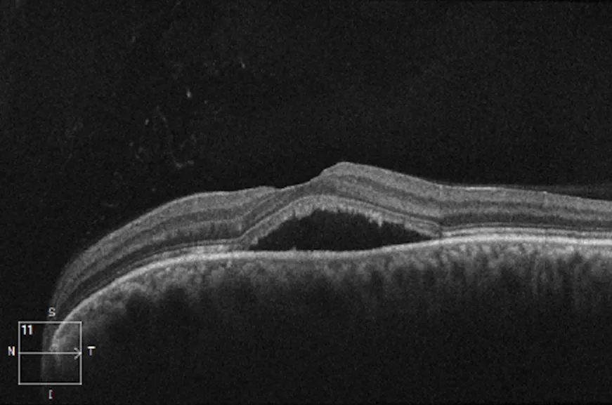
- 1Subretinal fluidAn optically empty (black) space between the neurosensory retina above and the RPE below: the retina has lifted off its pigment epithelium.
- 2Fovea, liftedThe foveal contour is preserved but the whole fovea sits on the dome of fluid, which is why the patient sees a grey smudge.
- 3Retinal pigment epitheliumThe bright continuous band. It is intact here; a break or a bump in it would point to a neovascular membrane instead.
- 4ChoroidThe mottled layer beneath the RPE. Thickened in central serous chorioretinopathy, which is part of why the fluid leaks.
Macular degeneration
Drusen are the first sign, deposits between the pigment epithelium and Bruch's membrane that show as pale yellow spots at the macula. Small hard drusen are common and mostly harmless; large soft confluent drusen and pigment clumping raise the risk of progression. Sudden distortion or a drop in vision in a patient with drusen means a new vessel until OCT proves otherwise, and it is seen within days because anti-VEGF injections work best early.
Examine it
Macular oedema, degeneration and holes take central vision; OCT shows them in cross section.
Ask OphthoSpace about the macula and fovea
AI responses are for education and may contain errors. OphthoSpace is not for primary clinical diagnosis.